WhatsApp
Neurosurgery:
Surgery of the Nervous System
Commonly referred to as Brain Surgery or Brain and Nerve Surgery, Neurosurgery is a comprehensive surgical discipline covering the entire nervous system.
3 Fundamental Pillars of the Nervous System:
- ● Brain: The control center.
- ● Spinal Cord: Communication bridge and protective spine.
- ● Nerves: Peripheral networks distributing to the body.
Which Diseases Are Treated?
Brain and Nerve Surgery manages the diagnosis and treatment of surgical conditions affecting the nervous system in both adults and children:
Vascular & Trauma
Vascular abnormalities, injuries, and hemorrhages.
Spine Health
Lumbar, cervical, thoracic herniations and canal stenosis.
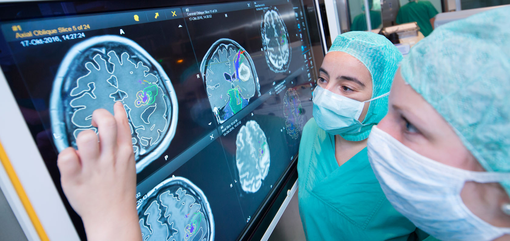
Oncological Surgery
Brain and spinal cord tumors.
Functional Surgery
Movement disorders and surgical nerve diseases.
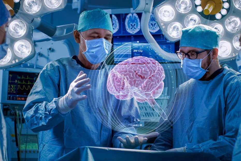
Academic Hospital Expertise
With our fully equipped expert staff, we utilize the latest techniques in treating central and peripheral nervous system diseases.
Spine and Spinal Cord Surgery
Pediatric Neurosurgery
Spine Surgery and Hernia Treatments
Lumbar Disc Herniation
Occurs when the cartilage tissue between the vertebrae protrudes outwards.
- ✓ Diagnosis: Confirmed via examination and MRI.
- ✓ Treatment: Rest, medication, and physical therapy are primary.
Microsurgery Method:
Cleaning is performed under a microscope through a small window. You can walk hours after surgery and be discharged the next day.
Cervical Disc Herniation
Causes pain and loss of strength in the arms. The hernia is removed via microsurgery and replaced with a prosthesis or cage. No risk of recurrence.
Spinal Stenosis
A condition that reduces quality of life and causes leg weakness. Surgery relieves the spinal cord, often using a screw system (plating) if necessary.
Pediatric Neurosurgery
Spina Bifida (Open & Closed)
Congenital spinal cord defect. Surgery within the first 24 hours is vital for open cases. Closed cases require MRI monitoring and timely intervention.
Hydrocephalus
Fluid accumulation in the brain. Treatment involves a Shunt (silicone tube) to drain excess fluid or endoscopic methods.
Early Intervention Saves Lives
In cases of brain hemorrhages, tumors, or nerve entrapments (Peripheral Nerve Surgery), early diagnosis is the key to success. At Academic Hospital, we safely treat your nervous system with microsurgery and modern imaging techniques.