
 WhatsApp
Diagnosis and Treatment Centre for Eye Health
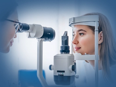
Ophthalmology is the medical specialty that diagnoses diseases affecting every structure of the eye and treats them with medical and surgical methods. Covering a broad spectrum that ranges from glaucoma to macular degeneration and from diabetic retinopathy to corneal disease, the department also manages systemic conditions that manifest in the eye, working in coordination with the diabetes treatment, neurology and internal medicine departments.
Contents
What Is the Ophthalmology Department?
The Ophthalmology department is responsible for the diagnosis and treatment of all diseases affecting the anterior segment (cornea, lens, iris) and the posterior segment (retina, vitreous, optic nerve) of the eye. Ophthalmologists manage medical treatment (eye drops, intravitreal injection) together with surgical treatment (cataract surgery, glaucoma surgery, retinal surgery). The department also plays a preventive role in avoiding vision loss through diabetic eye screening and regular fundus examinations.
Which Diseases and Conditions Are Treated?
The Ophthalmology department diagnoses and treats the following conditions and diseases:
- Cataract: Clouding of the lens of the eye; observation in the early stage and surgery by phacoemulsification (ultrasound) in the advanced stage
- Glaucoma (raised eye pressure): Management with drops, laser or surgery of optic nerve damage caused by increased intraocular pressure
- Age-related macular degeneration (AMD): Treatment with anti-VEGF injections when the centre of the retina (the macula) deteriorates with ageing
- Diabetic retinopathy: Screening for damage caused by diabetes to the retinal vessels, and treatment with laser photocoagulation and intravitreal injection
- Uveitis: Control of inflammation of the intraocular structures with steroid and immunomodulatory treatment
- Retinal detachment: Repair of the separation of the retina with laser and surgical methods; urgent assessment whenever it is suspected
- Vitreous haemorrhage: Management of bleeding into the vitreous due to diabetes, hypertension, trauma or vascular disease
- Corneal diseases: Medical and surgical treatment of keratoconus, corneal ulcer, herpetic keratitis and corneal dystrophies
- Dry eye syndrome: Treatment of the burning, stinging and blurred vision caused by insufficient tear production or tear quality
- Strabismus (squint): Correction of misalignment of the eyes with corrective lenses, patching therapy or surgical methods
- Amblyopia (lazy eye): Treatment of delayed visual development in childhood through early intervention
- Tinnitus and headache of ocular origin: Assessment of headaches related to raised eye pressure or refractive errors
- Eyelid diseases: Ptosis (drooping eyelid), entropion, ectropion and inflammatory eyelid disorders
When Should You Consult a Doctor?
The following symptoms call for an ophthalmological examination:
- Sudden or gradual reduction in visual acuity
- Flashes of light (photopsia) or seeing glare in the dark
- Darkening, a spot or a "curtain" sensation in the field of vision
- A sudden increase in the number of floaters
- Severe redness, pain or discharge in the eye
- Deterioration in night vision or seeing haloes (a sign of glaucoma)
- Drooping, swelling or a mass in the eyelid
- Double vision (diplopia)
Situations Requiring Emergency Attention
Emergency: The following situations require an emergency eye examination; permanent vision loss may occur within hours:
- Sudden loss of vision, or vision suddenly beginning to darken
- A curtain or shadow in the field of vision together with flashes of light (retinal detachment)
- Severe eye pain, disturbed vision, nausea and vomiting (acute angle-closure glaucoma)
- Entry of a chemical substance or a foreign body
- Blunt or penetrating trauma to the eye
Diagnostic Methods
In addition to the eye examination, advanced imaging and functional tests are used in the diagnosis of eye disease:
| Method | What it is used for |
|---|---|
| Detailed examination of the cornea, iris, lens and anterior segment | |
| The basic examination assessing the retina, optic nerve and macula; mandatory in the follow-up of diabetic retinopathy and glaucoma | |
| A non-invasive method that produces cross-sectional images of the retinal layers and the optic nerve at micron level; used in the follow-up of AMD, diabetic macular oedema and glaucoma | |
| Measures the central and peripheral field of vision in order to monitor the course of optic nerve damage of various causes | |
| The basic test for the diagnosis and follow-up of glaucoma | |
| Imaging of the retinal vessels with fluorescent dye to identify points of leakage and occlusion; used in AMD and diabetic retinopathy | |
| A test that maps the corneal surface; used in the diagnosis of keratoconus and in planning refractive surgery | |
| An advanced diagnostic method measuring the electrical response of the retinal cells | |
| Complementary tests assessing the quality of vision and colour perception |
Treatment Approaches
Treatment in ophthalmology involves planning medical, laser and surgical methods together according to the disease and its stage:
| Treatment | Scope and application |
|---|---|
| Beta blockers, prostaglandin analogues and carbonic anhydrase inhibitors that lower intraocular pressure in glaucoma; steroid and mydriatic drops in uveitis; artificial tears in dry eye | |
| Anti-VEGF agents (bevacizumab, ranibizumab, aflibercept) are injected into the eye in AMD and diabetic macular oedema | |
| Halting the formation of new vessels and sealing points of leakage in diabetic retinopathy | |
| Removal of the posterior capsule opacification (secondary cataract) that develops after cataract surgery | |
| Removal of the clouded lens by phacoemulsification (ultrasound) and implantation of an intraocular lens (IOL) | |
| Trabeculectomy or implantation of an intraocular shunt in cases that do not respond to medication and laser | |
| Vitreoretinal surgery with silicone oil or gas tamponade for the repair of detachment | |
| Partial or full replacement of the damaged cornea with advanced techniques (DSAEK, DALK) | |
| Eye muscle operations performed to correct the length and position of the eye muscles |
Book an appointment with the Ophthalmology Department.
Book an AppointmentOphthalmology at Academic Hospital
At the Academic Hospital Ophthalmology Department, outpatient examinations, advanced imaging investigations, laser applications and surgical procedures are carried out under one roof. The department works routinely with the following departments:
- Diabetes Treatment: joint management of diabetic retinopathy screening and blood glucose control
- Neurology: assessment of findings involving the optic nerve and the visual pathways
- Internal Medicine: initial assessment and referral of systemic diseases that manifest in the eye
- Endocrinology and Metabolic Diseases: follow-up of ocular findings related to diabetes and thyroid disease
- Rheumatology: joint follow-up of uveitis and ocular involvement in autoimmune disease
- Pediatrics: early screening and referral in amblyopia and strabismus
- Radiology: imaging investigations of the orbit and the visual pathways
Frequently Asked Questions
Answers to the questions most frequently asked about the Ophthalmology Department are given below.
References
The general information given on this page about eye diseases, diagnostic methods and treatment approaches is supported by the reliable health sources listed below.
- National Institutes of Health, "Open-Angle Glaucoma", StatPearls, PubMed Central (2024 update), ncbi.nlm.nih.gov
- Mayo Clinic, "Cataracts: Symptoms and Causes", Mayo Foundation (2024 update), mayoclinic.org
- World Health Organization, "Blindness and Visual Impairment", WHO (2024 update), who.int
- National Institutes of Health, "Diabetic Retinopathy", StatPearls, PubMed Central (2024 update), ncbi.nlm.nih.gov
The information on this page is for informational purposes only; it does not replace medical examination, diagnosis or treatment. Please consult a healthcare institution regarding your complaints.