
 WhatsApp
Minimally Invasive Procedures Under Imaging Guidance
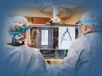
The Academic Hospital Interventional Radiology Unit performs minimally invasive procedures such as ultrasonography (US), computed tomography (CT) guided interventions and drainage. Because these procedures are carried out through small needle entries or millimetric incisions rather than open surgery, general anaesthesia is not required, the hospital stay is shorter and recovery is faster.
Contents
- What Is Interventional Radiology?
- In Which Diseases and Conditions Is an Interventional Approach Used?
- Interventional Procedures Performed
- Preparation Before the Procedure and the Process
- Which Departments Refer Patients to This Unit?
- Interventional Radiology at Academic Hospital
- Frequently Asked Questions
- References
What Is Interventional Radiology?
Interventional radiology procedures are carried out through smaller entry points than open surgery. As a result, patients feel less pain, stay in hospital for a shorter time and can return to daily life more quickly.
Interventional radiology is the medical speciality covering diagnostic and procedural interventions performed through very small incisions or needle entry points under the real time guidance of imaging methods such as X-ray, CT, US or DSA. Interventional radiologists are specialist physicians experienced both in imaging technology and in the use of instruments such as catheters, needles and ablation systems.
The unit works in close coordination with the imaging departments; procedure planning is based on CT and MRI findings, while the clinical decision rests on the request of the relevant specialist physician. Beyond producing a diagnosis on its own, it brings together tissue sampling under imaging guidance and minimally invasive intervention.
In Which Diseases and Conditions Is an Interventional Approach Used?
- Liver tumours (hepatocellular carcinoma and metastases): ablation
- Kidney tumours: percutaneous thermal ablation
- Lung tumours and suspicious lung nodules: CT guided biopsy and ablation
- Enlarged lymph nodes: percutaneous biopsy under imaging guidance
- Intra-abdominal and pelvic abscesses (collections of infected fluid): percutaneous drainage
- Pleural effusion (fluid around the lung) and ascites (fluid in the abdomen): drainage
Interventional Procedures Performed
Percutaneous Biopsy
A lung tissue sample is taken under CT guidance using a fine or core needle. The procedure is mostly completed within 60-90 minutes under local anaesthesia and the patient is discharged the same day. The tissue obtained is sent to the pathology department.
Abscess and Fluid Drainage
Intra-abdominal and fluid collections are drained through a thin drainage catheter placed under imaging guidance. This may remove the need for prolonged antibiotic treatment and for surgery.
Preparation Before the Procedure and the Process
| Stage | What happens |
|---|---|
| Blood clotting tests and existing imaging reports are reviewed. If you are taking blood thinning medication, it may need to be stopped for a period determined by your physician. | |
| For procedures requiring local anaesthesia, fasting of 3-4 hours is generally applied. | |
| The skin is cleaned and local anaesthesia is applied. The needle or catheter is guided to the target area under imaging. Most procedures are completed within 30-90 minutes. | |
| Depending on the type of procedure, patients are kept under observation for 2-6 hours; if no complication develops they are discharged the same day. |
Which Departments Refer Patients to This Unit?
Interventional radiology procedures are performed only at the request of a physician; patients cannot apply directly on their own:
- General Surgery: for abscess drainage and breast and thyroid biopsy.
Procedures at the Academic Hospital Interventional Radiology Unit are planned on physician referral. For an assessment and a procedure appointment you need a request document from your referring physician.
Get InformationInterventional Radiology at Academic Hospital
The Academic Hospital Interventional Radiology Unit works under the same roof as the imaging units; planning before the procedure, performing the intervention and follow-up afterwards are carried out within a single workflow. Tissue samples obtained are sent to the pathology department within the hospital. The unit works routinely with the following departments:
- General Surgery: requests for abscess drainage and breast and thyroid biopsy
- Radiology: the CT and MRI examinations that procedure planning is based on
- Medical Oncology: joint decisions on tumour ablation and diagnostic biopsy
- Pulmonology: suspicious lung nodules and pleural fluid interventions
- Pancreas, Liver and Bile Duct Diseases: biopsy and ablation in liver lesions
- Urology: percutaneous thermal ablation in kidney tumours
- Pathology: assessment of the tissue samples taken during the intervention
Frequently Asked Questions
References
The general information given on this page about interventional radiology methods, procedure workflows and patient preparation is supported by the reliable health sources listed below.
- Society of Interventional Radiology, "Patient Center: About Interventional Radiology", sirweb.org
- National Cancer Institute, "Types of Cancer Treatment", cancer.gov
- European Society of Radiology, "Patient Information", myesr.org
- Cardiovascular and Interventional Radiological Society of Europe, "Information for Patients", cirse.org
- Republic of Türkiye Ministry of Health, "Health Services Publications and Guidelines", dosyasb.saglik.gov.tr
The information on this page is for informational purposes only; it does not replace medical examination, diagnosis or treatment. Please consult a healthcare institution regarding your complaints.