

 WhatsApp
Definitive Diagnosis Through Tissue and Cell Analysis
Pathology is the branch of medicine that determines the origin, type, stage and spread of a disease by examining the structural changes occurring in tissues, organs and cells. The findings obtained in this process play a critical role in establishing the diagnosis and planning treatment.
Contents
- What Is Pathology?
- Which Examinations and Tests Are Performed in Pathology?
- In the Diagnosis of Which Diseases Does Pathology Play a Role?
- How Does the Analysis Process Work?
- How Long Do Results Take?
- How Is the Report Interpreted?
- Pathology at Academic Hospital
- Frequently Asked Questions
- References
What Is Pathology?
The pathology laboratory receives biopsy and surgical materials sent by a wide range of clinical departments, primarily the surgical units, endoscopy units, dermatology and radiology. Cytological samples such as Pap smears, most of which come from the obstetrics and gynaecology department, and other body fluids are also within the scope of assessment.
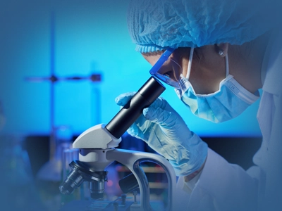
Sampling is carried out in the relevant clinical departments, and the material obtained is sent to the pathology laboratory in a suitable fixative that preserves the tissue. Every sample reaching the laboratory is first assessed macroscopically (with the naked eye) by the pathologist. Microscopic examination then follows tissue processing, thin sectioning and the staining procedures that make tissue and cellular changes recognisable. The pathological findings obtained are evaluated together with the clinical information and compiled into a standard pathology report. Pathology is therefore a discipline that plays a fundamental role in the diagnosis and treatment of disease and in shaping clinical decisions.
Which Examinations and Tests Are Performed in Pathology?
The pathology laboratory offers a broad range of diagnostic examinations; each method is used to answer a different clinical question.
- Routine histopathological examination: Tissue samples obtained by surgical, endoscopic or needle biopsy are processed in the laboratory, cut into thin sections, stained and assessed by the pathologist under the microscope. Cellular structure, the presence and type of tumour, the type of any inflammation and the tissue architecture are analysed.
- Intraoperative pathology consultation (frozen section): A tissue sample taken during surgery, while the patient is under anaesthesia, is rapidly frozen, sectioned, stained and examined under the microscope by the pathologist within a short time, and the result is reported to the surgeon. Delivered within minutes, this result helps the surgeon shape the operation.
- Cytopathological examinations (cytology):
- Fine needle aspiration cytology (FNA): Cellular material obtained with a fine needle from the thyroid, lymph nodes, breast and other masses is spread on a slide, stained and examined.
- Fluid cytology: The cellular content of body fluids such as pleural fluid collecting in the chest cavity, peritoneal fluid found in the abdominal cavity, cerebrospinal fluid and urine is assessed.
- Cervical cytology (Pap test / Pap smear): Cervical cells are screened for premalignant changes (changes that may progress to cancer).
- Immunohistochemical examination (immunohistochemistry - IHC): Specific antibodies are applied to tissue sections to assess whether the cells express particular proteins. It is used to determine the origin of a tumour, to establish hormone receptor status (such as ER, PR and HER2) and to identify treatment targets.
- Molecular pathology examinations: The process of detecting genetic changes in the DNA and RNA within the cell.
- FISH (fluorescence in situ hybridisation): Used to detect gene amplifications and chromosomal rearrangements. It has a particular place in assessing targets such as HER2 status.
- PCR-based molecular tests: These allow gene mutations such as EGFR, KRAS and BRAF to be detected and play an important role in the choice of targeted therapy.
- Bone marrow aspiration and biopsy examination: Bone marrow samples sent by the haematology department are assessed, contributing to the diagnosis of haematological diseases such as leukaemia, lymphoma and myeloma.
In the Diagnosis of Which Diseases Does Pathology Play a Role?
Pathology plays a fundamental role in establishing the definitive diagnosis in many diseases and in determining their type, stage and biological behaviour. It is indispensable in particular where assessment at tissue and cell level is required.
- Tumours and cancers: Distinction between benign and malignant tumours, tumour type, how far the tumour cell has departed from normal, its grade and its extent (staging)
- Gastrointestinal system diseases: H. pylori gastritis, inflammatory bowel diseases (ulcerative colitis, Crohn's disease), coeliac disease, polyps and malignancies
- Liver diseases: Activity and fibrosis staging in the various types of hepatitis, cirrhosis, fatty liver disease and liver tumours
- Autoimmune diseases: Conditions in which tissue involvement is assessed, such as lupus nephritis (kidney biopsy) and myositis (muscle biopsy)
- Skin diseases: Melanoma and other skin tumours, psoriasis and vasculitis
- Haematological diseases: Leukaemia, lymphoma and bone marrow failure (for example aplastic anaemia)
- Endocrine and solid organ tumours: Thyroid, breast, lung and prostate tumours
- Diseases of the female genital system: Cervical dysplasia, endometrial polyps, hyperplasia and cancers, ovarian malignancies
- Transplantation medicine: Assessment of rejection in organ transplants such as kidney and liver
How Does the Analysis Process Work?
The pathology analysis process is a standardised workflow, managed with quality control procedures, that covers every stage from acceptance of the sample into the laboratory to preparation of the pathological diagnosis report. The process is critically important for accurate diagnosis, traceability and patient safety.
| Stage | What happens |
|---|---|
| Surgical material, biopsy and cytology samples sent from surgical units, endoscopy units, outpatient clinics or other clinical areas arrive at the laboratory. Samples are registered in the system together with patient identity verification, barcoding, sample type and preliminary clinical diagnosis details. | |
| Samples are assessed by the pathologist with the naked eye. Tissue size, weight, colour, surface characteristics and the description of the lesion are recorded. Diagnostically important areas are identified, sampling is carried out and the material is placed in tissue cassettes. | |
| Tissue samples are fixed in a suitable fixative (usually formalin). They then pass through dehydration, clearing and paraffin infiltration to harden the tissue. Once the paraffin blocks containing the tissue have been prepared, sections 3-5 microns thick (1 mm = 1000 microns) are cut with a microtome and transferred onto glass slides. | |
| Haematoxylin-Eosin (H&E) staining is applied routinely. Where necessary for an accurate diagnosis, additional pathological assessments are made on the sections prepared in the laboratory using special histochemical stains and immunohistochemical (IHC) methods. | |
| The pathologist examines the prepared slides under the microscope in the light of the patient's clinical information. The morphological findings in the tissue and cells are assessed and concluded. The findings are interpreted together with the clinical information and international classification systems, and the pathology report is produced. | |
| The histopathological diagnosis and the pathology report are prepared in a format that complies with the applicable standards. After passing through the department's quality control stages, the report is sent to the requesting physician through the electronic system. |
How Long Do Results Take?
Turnaround times vary according to the type of examination.
| Type of examination | Average turnaround time |
|---|---|
| An average of 3-5 working days | |
| An average of 3-5 working days | |
| 10-20 minutes | |
| An average of 2-3 working days is added to the routine biopsy time |
In emergencies, or in cases awaiting an oncological decision, a priority examination protocol is applied.
How Is the Report Interpreted?
The pathology report is one of the most important medical documents in the diagnostic process. Correct interpretation of the report is carried out by the treating clinician, who evaluates it together with the patient's clinical findings, imaging results and other laboratory data. Where necessary, the clinical and pathology departments meet to discuss the patient's condition.
Pathology reports generally state the organ and anatomical location the sample came from, the macroscopic findings (size and appearance), the microscopic assessment, the histopathological diagnosis and, where applicable, the pathological staging (such as pT and pN). In cancer cases the report also states prognostic features such as the histological type and grade of the tumour, the surgical margin status and lymphovascular invasion.
The term "malignant" refers to malignant tumours (cancer) and "benign" to benign lesions. "Dysplasia" indicates disruption in cell structure and organisation, a finding that in some cases may carry premalignant potential.
A pathology report is not merely a diagnostic document; it is also a medical document that plays a fundamental role in shaping the treatment plan. The surgical approach, drug therapy and follow-up strategies are largely determined by this report.
Important: For this reason, interpreting a pathology report on your own or over the internet is not appropriate. Results should only be explained by the treating physician, assessed together with the clinical picture and the other findings.
Get information about the Academic Hospital Pathology Laboratory.
Get InformationPathology at Academic Hospital
The Academic Hospital Medical Pathology Laboratory assesses tissue and cell samples from the hospital's clinical departments under one roof; frozen section consultations requested during surgery are also carried out in this laboratory. The laboratory works routinely with the following departments:
- Medical Oncology: treatment planning based on tumour type, grade and biological markers
- General Surgery: assessment of surgical materials and intraoperative consultation
- Endoscopy: examination of gastric, intestinal and upper digestive system biopsies
- Dermatology: assessment of biopsies taken from skin lesions
- Radiology: examination of needle biopsies taken under imaging guidance
- Gynecology and Obstetrics: cytopathological examination of Pap smears and gynaecological materials
- Hematology: assessment of bone marrow aspiration and biopsy samples
Frequently Asked Questions
References
The general information given on this page about pathological examination methods, the analysis process and reporting standards is supported by the reliable health sources listed below.
- College of American Pathologists, "CAP Cancer Protocols and Checklists", cap.org
- Royal College of Pathologists, "Histopathology Dataset and Tissue Pathway Guidelines", rcpath.org
- European Society of Pathology, "ESP Guidelines and Quality Assurance", esp-pathology.org
The information on this page is for informational purposes only; it does not replace medical examination, diagnosis or treatment. Please consult a healthcare institution regarding your complaints.
Choose a Doctor