

 WhatsApp
A Bridge to Diagnosis Through Imaging
The Academic Hospital Radiology Department examines the internal structures of the body with high-resolution imaging technologies, providing clinicians with accurate and timely diagnostic support. The department does not deliver treatment; radiology specialists interpret the images they produce and send a written report to the referring physician. A broad range of services is offered, including X-ray, ultrasound, computed tomography (CT), magnetic resonance imaging (MRI), mammography and bone densitometry.
Contents
What Is Radiology?
Radiology is the medical specialty that images the internal structures of the body using physical energy sources such as X-rays, sound waves, magnetic fields and radioactive tracers. Specialist radiologists interpret these images and contribute to the detection and staging of disease and to monitoring the response to treatment. Alongside diagnostic radiology, interventional procedures such as image-guided biopsy or drainage are carried out by the Academic Hospital Interventional Radiology Department. The treatment decision always rests with the relevant clinical specialist.
Which Conditions Does It Help to Diagnose?
The radiology department does not treat disease; it provides the clinician with imaging evidence for the diagnosis. The clinical scenarios for which imaging is most often requested are the following:
- Follow-up of lung nodules, pneumonia and staging of lung cancer
- Assessment of cerebral haemorrhage, stroke and intracranial masses
- Preoperative anatomical mapping in gastric, colon or pancreatic cancer
- Coronary CT angiography where coronary artery stenosis is suspected
- Detection and characterisation of liver, pancreatic and kidney masses
- Joint cartilage damage, meniscal tears and disc herniation
- Ultrasound of thyroid nodules and neck masses
- Breast masses, microcalcifications and breast cancer screening
- Bone mineral density measurement for osteoporosis risk assessment
- Investigation of internal organ injury after acute abdomen or trauma
- Follow-up hip ultrasound in newborns at two months of age
Imaging Methods and Technologies Used
Each imaging method visualises different tissue types with different sensitivity; the clinician decides which method will be used.
Plain Radiography (X-ray)
Plain radiography (X-ray) is a basic imaging method that allows the internal structures of the body to be visualised rapidly using X-rays. It is frequently used as a first-line examination, particularly in the assessment of bone fractures, lung disease and heart size. It can also be helpful in the assessment of some abdominal conditions. Because it can be performed quickly, is widely available and takes only a short time, it is one of the most frequently used radiological methods.
Ultrasonography (Ultrasound)
Ultrasonography is an imaging method that creates real-time images using sound waves and involves no ionising radiation. It is widely used in the assessment of the liver, gallbladder, kidneys and other abdominal organs, as well as the thyroid, breast, muscle and tendon structures and blood vessels. Because it involves no ionising radiation, it is often the first-choice imaging method in children and pregnant women. Doppler ultrasound also allows the structure of vessels and blood flow to be assessed.
Computed Tomography (CT)
Computed tomography (CT) is an imaging method that produces cross-sectional images of the body using X-rays and computer technology. It provides rapid and detailed assessment in acute trauma, lung and abdominal disease, vascular pathology and cancer staging. Where required, a contrast agent can be used so that vascular structures and organs can be assessed more clearly.
Coronary CT Angiography
By imaging the heart and the coronary arteries at high resolution, this method allows arterial stenosis to be assessed non-invasively. It provides detailed information on coronary artery disease and, in suitable patients, may reduce the need for invasive (catheter) angiography.
Magnetic Resonance Imaging (MRI)
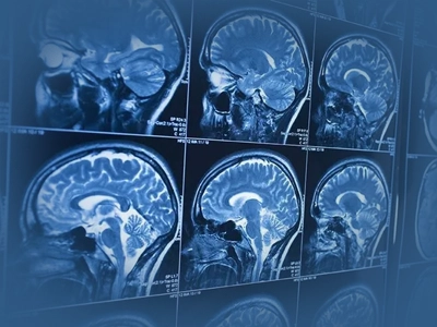
Magnetic resonance imaging (MRI) is a method that allows detailed imaging of the soft tissues of the body using a magnetic field and radio waves. It involves no ionising radiation. Thanks to its high soft-tissue contrast, it provides important information in the assessment of the brain, spinal cord, joints, musculoskeletal system and pelvic organs.
Mammography
Mammography is a method that images breast tissue using low-dose X-rays. It plays an important role in the screening and diagnosis of breast cancer. Digital mammography and, where required, 3D tomosynthesis can help detect small lesions and early changes.
Bone Densitometry (DEXA)
Bone densitometry (DEXA) measures bone mineral density in the spine and hip region, helping to establish a diagnosis of osteoporosis and contributing to the assessment of fracture risk. It is frequently used to assess bone health, particularly in postmenopausal women and in patients on long-term corticosteroid treatment.
When and How Are Results Ready?
The time needed to prepare reports for radiological examinations may vary according to the type of examination, its technical characteristics and the level of urgency.
| Examination | Reporting time |
|---|---|
| The same day or within 1 working day | |
| Usually shortly after the procedure or the same day | |
| Routine: 1-2 working days; urgent: within 30-60 minutes | |
| Usually 1-3 working days | |
| 1-2 working days | |
| After 1 hour |
Reports are sent to the requesting physicians through the digital system. To obtain information about your results you should speak to the doctor who referred you; the radiology department does not provide direct patient consultations or interpret results for patients.
Which Departments Refer Patients to Radiology?
Radiology is a central diagnostic infrastructure serving every clinic; examinations are always requested by a specialist physician.
- Cardiology: coronary CT angiography
- Medical Oncology: mass staging and monitoring of treatment response
- Neurology and Neurosurgery: brain and spinal cord MRI, cranial CT
- Orthopaedics and Traumatology: bone radiographs, joint MRI, trauma CT
- General Surgery and Gastroenterology: ultrasound, CT and MRI of the abdominal organs
- Gynecology and Obstetrics: pelvic ultrasound, breast ultrasound
- Endocrinology: thyroid ultrasound, adrenal CT, adrenal MRI and pituitary MRI
- Emergency Department: priority imaging for trauma, stroke and acute abdomen protocols
Book an appointment with the Academic Hospital Radiology Department.
Book an AppointmentRadiology at Academic Hospital
The Academic Hospital Radiology Department handles imaging requests from the hospital's outpatient clinics, wards and emergency areas in a single centre; reports are sent to the requesting physician through the digital system. The department works routinely with the following units:
- Cardiology: assessment of arterial stenosis with coronary CT angiography
- Medical Oncology: mass staging and imaging follow-up of treatment response
- Neurology: brain and spinal cord MRI and cranial CT examinations
- Orthopaedics and Traumatology: bone radiographs, joint MRI and trauma CT
- General Surgery: preoperative anatomical mapping and imaging of the abdominal organs
- Breast Diseases and Surgery: mammography and breast ultrasound assessments
- Interventional Radiology: image-guided biopsy and drainage procedures
Frequently Asked Questions
References
The general information given on this page about imaging methods, radiation safety and reporting processes is supported by the reliable health sources listed below.
- Radiological Society of North America, "RadiologyInfo.org: Mammography", radiologyinfo.org
- International Atomic Energy Agency (IAEA), "Radiation Protection in Radiology", iaea.org
- Mayo Clinic, "MRI: Overview, Purpose and What to Expect", mayoclinic.org
- NHS, "CT Scan: What It Is and When It Is Used", nhs.uk
- World Health Organization (WHO), "Cancer: Key Facts", who.int
The information on this page is for informational purposes only; it does not replace medical examination, diagnosis or treatment. Please consult a healthcare institution regarding your complaints.