Diagnosis and Treatment in Chronic Venous Insufficiency and Leg Varicose Veins
Published:
· Last Updated:
· Prepared by the Academic Hospital Web and Editorial Board.
The Varicose Vein Treatment Unit carries out the diagnosis and treatment of venous disease, from valve failure in the leg veins to fine spider veins. Assessment begins with detailed vein mapping using colour Doppler ultrasound, and the treatment plan is built for each person according to the underlying cause of the disease.
The aim of varicose vein treatment is not only to remove the visible veins, but first to identify and treat the underlying problem in the venous system. For this reason the leg veins are always assessed with colour Doppler ultrasound before treatment begins.
During the examination, the diameter of the great and small saphenous veins, the presence of leakage in the vein valves (venous reflux), the patency of the deep venous system and the reticular veins that feed the spider veins are all examined in detail.
If valve failure is found in the large veins, this problem is treated first with methods such as endovenous laser (EVLA), radiofrequency ablation (RFA), venous adhesive or, in suitable patients, foam sclerotherapy. Treating only the spider veins without correcting the underlying reflux may lead to new spider veins within a short time and to repeated treatment.
Once the leakage in the venous system has been treated, microsclerotherapy, electrothermocoagulation and Nd:YAG laser treatments can be applied alone or in combination for the remaining cosmetic spider veins. Which method is used is decided individually according to the diameter, colour, depth and location of the vein and the patient's skin structure.
The basic principle of successful and lasting varicose vein treatment is to treat the cause first and then to remove the visible veins for a cosmetic result.
What Are Varicose Veins?
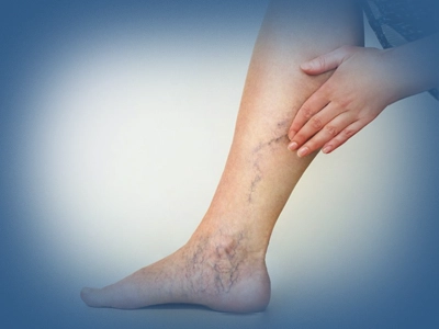
Varicose veins are a common vascular disease that appears when the veins in the legs widen and become prominent. Normally the valves inside the veins allow blood to flow in one direction, from the leg towards the heart, against gravity. When these valves can no longer do their job (valve failure or venous reflux) the blood flows back down into the legs and pools inside the vein. Over time the vein widens, takes on a tortuous appearance and a varicose vein forms. This is not only a cosmetic problem; it can cause pain, a feeling of heaviness, burning, night cramps, swelling (oedema) and itching in the legs, and in later stages skin colour changes and even ulcers. A family history of varicose veins, working for long periods standing or sitting, pregnancy, excess weight, advancing age and a sedentary lifestyle are the most important factors that increase the risk of developing varicose veins.
Which Diseases and Conditions Are Treated?
Varicose veins and venous diseases are not only a cosmetic problem. With early diagnosis and appropriate treatment, symptoms can be reduced and progression of the disease can be prevented. The diagnosis and treatment of the following venous diseases are carried out in our clinic:
Spider veins (telangiectasia): fine red, purple or spider web shaped vessels seen on the skin surface.
Reticular varicose veins: medium calibre superficial veins of a blue and green colour seen under the skin.
Insufficiency and varicose veins of the great saphenous vein (vena saphena magna).
Insufficiency and varicose veins of the small saphenous vein (vena saphena parva).
Chronic venous insufficiency: venous disease that runs with pain, a feeling of heaviness, fatigue, swelling (oedema), burning and night cramps in the legs.
Venous stasis dermatitis: skin colour change, itching and eczema like findings around the ankle caused by venous insufficiency.
Lipodermatosclerosis: hardening and brown discolouration of the skin and subcutaneous tissue as a result of long standing venous insufficiency.
Venous ulcer: open wounds around the ankle caused by venous insufficiency that heal slowly.
Superficial thrombophlebitis: the development of a clot and vein inflammation in superficial varicose veins.
Pregnancy related varicose veins: assessment after delivery and treatment at the appropriate time of varicose veins that appear or become prominent during pregnancy.
Recurrent varicose veins: assessment and treatment of varicose veins that form again although they have been treated before.
When Should You Consult Us?
If you have one or several of the following symptoms, it is recommended that you consult a cardiovascular surgery specialist:
A marked feeling of heaviness, fullness and fatigue in the legs at the end of the day
Swelling (oedema) of the ankles and legs, particularly after standing for a long time
Leg cramps that appear at night
Burning, aching, itching or a feeling of restlessness in the legs
Spider veins or varicose veins on the skin surface becoming gradually more prominent
The formation of tortuous and widened veins in the leg
Darkening of the skin colour, hardening or eczema like changes around the ankle
The formation of slowly healing or recurrent wounds (venous ulcers) on the leg
The development of new varicose veins although varicose vein treatment has been carried out before
Emergencies: If the following symptoms appear, the emergency medical service should be called without delay or the nearest emergency department should be attended:
Sudden severe swelling, pain, redness and increased warmth in the leg
Painful swelling along a hard and tender vein (superficial thrombophlebitis)
Rapidly developing swelling and pain in one leg (suspected deep vein thrombosis, DVT)
If these symptoms are accompanied by sudden shortness of breath, chest pain, palpitations, fainting or blood stained sputum, a pulmonary embolism (a clot travelling to the lung) may have developed. Because this condition can be life threatening, it requires urgent medical assessment.
Diagnostic Methods
Detailed Vein Mapping with Colour Doppler Ultrasound
The first and most important step of varicose vein treatment is the detailed assessment of the leg venous system with colour Doppler ultrasound. During the examination the study is most often performed by the cardiovascular surgery specialist, with the patient standing and in a dynamic manner. When needed, or in complex cases, Doppler ultrasound may be repeated by a radiology specialist in order to support the assessment.
This examination produces what is effectively a map of the leg veins. The diameter of the great and small saphenous veins, the presence of leakage in the vein valves (venous reflux), the patency of the deep venous system, the perforating (connecting) veins, the reticular veins that feed the spider veins and findings of any previous clot or vein occlusion are all assessed in detail.
Thanks to this mapping, the real cause of the patient's varicose veins is identified and the most appropriate treatment plan is drawn up. Unnecessary interventions are thus avoided, the correct vein is treated, and long term success and patient satisfaction increase.
CT or MR venography: Not frequently used. When deep vein disease or pelvic venous insufficiency is suspected, the vein anatomy is assessed in detail with cross sectional imaging.
Treatment Approaches
The aim of varicose vein treatment is not only to remove the visible veins, but first to detect and treat venous valve failure (venous reflux), the real cause of the disease. For this reason, before treatment the diameter of the great and small saphenous veins, the presence of leakage in the vein valves (venous reflux), the patency of the deep venous system and the veins that feed the spider veins are always assessed in detail with colour Doppler ultrasound. If valve failure is found in the large veins, this problem is treated first with methods such as endovenous laser ablation (EVLA), radiofrequency ablation (RFA), venous adhesive or, in suitable patients, foam sclerotherapy. Treating only the spider veins without correcting the underlying venous reflux may lead to the formation of new varicose veins and to repeated treatment within a short time.
Today the methods most frequently used in the treatment of large vein varicosities are endovenous laser (EVLA), radiofrequency ablation (RFA) and venous adhesive treatment. The shared aim of these methods is to close the vein with valve failure from the inside so that blood is directed to healthy veins. The success rate of all three methods is above 90 to 95 per cent, and they are among the first line treatment options in international guidelines.
Method
Scope and application
In EVLA a thin laser fibre is placed inside the vein and the vein is closed from the inside using laser energy. It is particularly effective in wide calibre saphenous veins. The long term success rate is high and it is one of the most widely applied methods in the world. The procedure is performed under local anaesthesia and patients can walk home on the same day.
In radiofrequency treatment, radiofrequency energy that produces controlled heat is used instead of laser. Because the vein wall is heated more evenly, bruising and pain after the procedure may be slightly less in some patients. Success rates are similar to EVLA and it is again applied under local anaesthesia.
In this method a special medical adhesive is delivered inside the vein to close it. Because heat is not used, tumescent anaesthesia (numerous local anaesthetic injections around the vein) is not required. After the procedure many patients may not need to wear compression stockings and the return to daily life is quite rapid. It is an important option particularly for patients who wish to reduce the number of needles.
Which method is better?
No single method is the best option for all patients. The choice of treatment is determined by the diameter of the vein, the structure of the vein, the site of the leakage (venous reflux), the patient's expectations, accompanying diseases and the surgeon's experience. According to current international guidelines, the long term success rates of EVLA, RFA and venous adhesive treatments are quite close to one another in suitable patients. The most important element is applying the right method to the right patient.
Cosmetic Varicose Vein Treatments: Microsclerotherapy, Electrothermocoagulation and Nd:YAG Laser
In the treatment of spider veins, no single method is the most appropriate option for every patient. An individual treatment plan is drawn up after assessing the diameter, colour, depth and location of the vein and the skin type. For this purpose microsclerotherapy, electrothermocoagulation and Nd:YAG laser treatments can be applied alone, or combined in the same patient in order to obtain the best cosmetic result.
What Is Spider Vein Treatment (Microsclerotherapy)?
Microsclerotherapy is one of the most frequently used methods with scientifically proven effectiveness in the treatment of the fine purple, red or blue spider veins seen on the legs (telangiectasia and reticular veins). During the procedure a special medication (a sclerosant agent) is injected into the spider veins with very fine needles. This medication creates a controlled reaction in the vein wall and causes the vein to close. Over time the treated vein is absorbed by the body and its visibility disappears.
The procedure is usually performed in outpatient conditions, does not require anaesthesia and takes about 15 to 30 minutes. After treatment the patient can walk out and return to daily life. Short term use of compression stockings may be recommended in order to obtain a better result. More than one session may be needed depending on how widespread the spider veins are.
Before microsclerotherapy is applied, underlying venous insufficiency must always be investigated with Doppler ultrasound. This is because if there is valve failure in the large veins, the risk of recurrence is higher in spider vein treatments carried out without first treating that problem.
After treatment, mild bruising, redness or firmness along the vein may be seen for a few weeks. In most patients these resolve on their own. When the patient is suitably selected, microsclerotherapy is a safe and effective treatment method that both improves the cosmetic appearance and provides high patient satisfaction.
Electrothermocoagulation is the procedure of closing a spider vein from the inside by delivering high frequency microwave energy into the vein through a very fine probe. It is an effective option particularly for superficial, very fine (under 0.3 mm) red spider veins, resistant veins around the ankle and small veins that are difficult to treat with a needle.
Nd:YAG laser (1064 nm) is a laser treatment applied through the skin that targets the haemoglobin inside the vein and closes it. It may be preferred in patients who do not want needles, and in the treatment of the face, the ankle or certain fine spider veins. It can also be effective on veins that lie deeper and are slightly thicker.
In many patients the most successful result is obtained by using these methods together. For example, while thicker reticular veins are treated with microsclerotherapy, the very fine red spider veins that remain can be completed with electrothermocoagulation or Nd:YAG laser. In this way the cosmetic result improves and treatment success increases.
Before treatment begins, underlying venous valve failure must always be investigated with colour Doppler ultrasound. If there is venous reflux in the large veins, treating that disease first reduces the risk of the spider veins recurring. The treatment method, or the combination of methods, should be planned individually in line with the patient's vein structure and expectations.
For deep vein disease or varicose veins of pelvic origin, a coordinated assessment is made with the Cardiovascular Surgery Unit. Joint management is provided with the internal medicine teams for wound care and with the cardiology teams for long term follow up. In the choice of treatment method, the patient's age, general state of health, the findings of the venous mapping and the patient's preferences are all assessed together. In some situations a combination of more than one method may give the best result; this decision is individualised through a multidisciplinary approach.
Vein mapping with colour Doppler ultrasound, closed vein treatments and cosmetic spider vein treatments are provided in a single centre at the Academic Hospital Varicose Vein Treatment Unit. Tel: 444 0 353
The Academic Hospital Varicose Vein Treatment Unit carries out outpatient assessment, vein mapping with colour Doppler ultrasound, closed vein treatments and cosmetic spider vein procedures under one roof. The unit works routinely with the following departments:
Cardiovascular Surgery: coordinated assessment of deep vein disease and varicose veins of pelvic origin
Radiology: repeat Doppler ultrasound and cross sectional venography in complex cases
Emergency Department: urgent assessment when deep vein thrombosis or pulmonary embolism is suspected
Frequently Asked Questions
Is varicose vein treatment painful?
No. The modern varicose vein treatments used today are in general mildly painful or almost painless procedures. Endovenous laser (EVLA), radiofrequency (RFA) and venous adhesive treatments are applied under local anaesthesia and the great majority of patients describe only a mild pressure or needle sensation during the procedure. In microsclerotherapy and spider vein treatments, very fine needles are used, so the discomfort that occurs is usually short lived and mild. After the procedure, mild tightness, bruising or tenderness that can last a few days may be seen, and most patients can walk out and return to daily life on the same day. Although the level of pain can vary with the treatment method used, varicose vein treatments today are tolerated comfortably by the great majority of patients.
Is surgery necessary for varicose vein treatment?
Not every patient with varicose veins needs surgery. Today a significant proportion of large vein varicosities can be treated successfully with closed (minimally invasive) treatment methods applied under the guidance of colour Doppler ultrasound. The methods most frequently used include endovenous laser (EVLA), radiofrequency ablation (RFA) and venous adhesive treatments. In these procedures no large incision is made in the groin or the leg; a thin catheter is placed inside the vein under ultrasound guidance and the vein with valve failure is closed from the inside. The procedure is carried out under local anaesthesia, usually takes 30 to 60 minutes, and patients can walk home on the same day. Today classical open varicose vein operations are preferred only in a limited number of patients for whom closed methods are not suitable. Which treatment is most appropriate for you is decided after a detailed assessment with colour Doppler ultrasound.
Can I walk on the same day after varicose vein treatment?
Yes. After the modern varicose vein treatments applied today, namely endovenous laser (EVLA), radiofrequency (RFA), venous adhesive, foam sclerotherapy and microsclerotherapy, the great majority of patients can stand up and walk on the same day. Indeed, taking short walks is particularly recommended because it increases blood circulation in the leg veins and reduces the risk of clot formation. A walk of about 20 to 30 minutes is usually advised after the procedure. Most patients can return to daily life the same day; however, heavy sport, intense exercise and lifting heavy loads should be avoided for the first few days. Your doctor will give you individual advice on the use of compression stockings according to the treatment method used.
When can I return to work after varicose vein treatment?
Although this period varies with the treatment method used and with your work, after the modern varicose vein treatments applied today the great majority of patients can return to daily life and to desk work on the same day or the next day. With minimally invasive methods such as endovenous laser (EVLA), radiofrequency (RFA), venous adhesive, foam sclerotherapy and microsclerotherapy, a hospital stay is not required and recovery is quite rapid. People who work in jobs requiring heavy physical effort are generally advised to avoid heavy lifting and activities requiring intense effort for 3 to 7 days. Your doctor will give you the most appropriate advice about your return to work according to the treatment applied and your occupation.
Do varicose veins recur after treatment?
After successful varicose vein treatment it is rare for the treated vein to open again, but because varicose vein disease is a chronic and progressive venous disease, new varicose veins may develop in different veins over the years. The risk of recurrence depends on genetic predisposition, working for long periods standing, pregnancy, excess weight, a sedentary life and how widespread the underlying venous valve failure is. For this reason, varicose vein treatment must address not only the visible veins but also the valve failure detected with colour Doppler ultrasound. Through regular check ups, healthy lifestyle habits and preventive treatment where necessary, the risk of new varicose veins forming can be reduced considerably.
Do spider veins disappear completely?
In most patients spider veins are markedly reduced with appropriate treatment and a large proportion of them can disappear completely. However, the result depends on the diameter of the veins, how widespread they are, the skin structure and the presence of underlying venous disease. In some patients more than one treatment session may be needed to reach the best cosmetic result. In addition, if there is valve failure (venous reflux) in the large veins that feed the spider veins, that problem must be treated first. Otherwise new spider veins may develop. Today, quite successful cosmetic results can be obtained by applying microsclerotherapy, electrothermocoagulation and Nd:YAG laser treatments alone or in combination. Most disappear completely, and for some an additional session may be needed.
How many sessions of microsclerotherapy are applied?
The number of sessions needed for microsclerotherapy varies with how widespread the spider veins are, the diameter of the veins and the size of the area to be treated. In most patients 2 to 4 sessions are sufficient, while people with widespread spider veins may need 5 or more sessions. Sessions are usually planned 3 to 6 weeks apart. Although a large number of veins can be treated in the same session, staged treatment is preferred for the best cosmetic result and for safety. In addition, if there is valve failure (venous reflux) in the large veins, treating that problem first increases the success of microsclerotherapy and reduces the risk of new spider veins forming.
Who is foam treatment (foam sclerotherapy) applied to?
Foam sclerotherapy is an effective treatment method that can be applied particularly in patients who have medium and large calibre superficial varicose veins, who have venous valve failure, or who are not suitable for surgery or endovenous treatment. During the procedure a special medication prepared under ultrasound guidance is turned into foam and injected into the varicose vein. The foam affects the vein wall, causing the vein to close and in time to be absorbed by the body. It can be used successfully in selected patients with valve failure in the great or small saphenous vein, in side branch varicosities, in recurrent varicose veins and in the treatment of varicose veins remaining after surgery. The treatment decision is made after a detailed assessment with colour Doppler ultrasound, according to the vein structure and the patient's characteristics.
Who is foam sclerotherapy not applied to?
Foam sclerotherapy is not suitable for every patient with varicose veins. It is not applied during pregnancy or in patients with active deep vein thrombosis (DVT) or a recent venous clot. In people with a known allergy to the medication to be used, in patients with an active infection in the area to be treated and in people with a serious uncontrolled systemic disease, treatment may also be postponed or different methods may be preferred. In advanced peripheral arterial disease, in patients who are unable to walk because of severe immobility and, in certain selected situations, in people with a right to left cardiac shunt (particularly a symptomatic patent foramen ovale, PFO), a detailed assessment is required before the procedure. In addition, in patients with marked valve failure in the large veins, whether foam treatment alone will be sufficient is assessed with colour Doppler ultrasound. The most appropriate treatment method is determined individually according to the patient's vein structure, general state of health and ultrasound findings.
What is the difference between endovenous laser ablation (EVLA) and radiofrequency ablation (RFA)?
Ablation is used in the sense of burning and closing. Endovenous laser (EVLA) and radiofrequency ablation (RFA) are two very similar modern closed treatment methods used in the treatment of venous valve failure. Both procedures are applied under the guidance of colour Doppler ultrasound and under local anaesthesia, and the aim is to close the vein with valve failure from the inside and direct blood flow to healthy veins. The most important difference is the energy source used to close the vein. EVLA uses laser energy, while RFA closes the vein by heating the vein wall with controlled radiofrequency energy. The long term success rate of both methods is above 90 to 95 per cent and they are among the first line treatment options in international guidelines. While some studies report slightly less pain and bruising after RFA, this difference has largely been reduced with new generation laser systems. Which method is more appropriate is determined by the diameter of the vein, its anatomical structure, the patient's characteristics and the physician's experience. Today both methods are safe and effective treatments that allow discharge on the same day.
Who is venous adhesive treatment suitable for?
Venous adhesive treatment is a modern, minimally invasive varicose vein treatment applied particularly in patients with valve failure (venous reflux) in the great or small saphenous vein. In this method a thin catheter is placed inside the vein and a special medical adhesive is delivered under ultrasound guidance to close the diseased vein. Because heat is not used, there is no need for numerous local anaesthetic injections around the vein (tumescent anaesthesia). It may be an important option for patients who are wary of needles, who do not wish to wear compression stockings, or who want to return to daily life and work as quickly as possible. However, not every patient is suitable for this method. The treatment decision is made individually after the diameter of the vein, the site of the valve failure, the vein structure and the patient's general state of health have been assessed with colour Doppler ultrasound.
Which of EVLA, RFA and venous adhesive treatments is better?
No single method is the best option for all patients. Endovenous laser (EVLA), radiofrequency ablation (RFA) and venous adhesive treatments are all modern methods with a high success rate that current international guidelines recommend for the treatment of valve failure in the large veins. The choice of treatment is determined by the diameter of the vein, the vein structure, how widespread the valve failure is, the patient's general state of health and expectations, and the physician's experience. In EVLA and RFA the vein is closed with heat energy and local anaesthesia (tumescent anaesthesia) is applied around the vein. In venous adhesive treatment, because heat is not used, tumescent anaesthesia is not required and some patients may not need to wear compression stockings. Today the long term success rates of all three methods are quite close to one another. The most successful result is obtained by choosing the right method for the right patient after a detailed assessment with colour Doppler ultrasound.
Is it necessary to wear compression stockings after varicose vein treatment?
This depends on the treatment method used. After endovenous laser (EVLA), radiofrequency ablation (RFA) and foam sclerotherapy, your doctor may advise you to wear compression stockings for a certain period in order to support healing and reduce bruising and tenderness. By contrast, in venous adhesive treatment the use of compression stockings is not obligatory for many patients. However, this decision may vary according to the characteristics of the treated vein, any additional procedures performed and the patient's general condition. The most appropriate duration and stocking pressure are determined individually by the physician performing the procedure.
Can I do sport after varicose vein treatment?
Yes. However, the time of return to sport may vary with the treatment method used. After endovenous laser (EVLA), radiofrequency (RFA), venous adhesive, foam sclerotherapy and microsclerotherapy, taking a short walk on the same day is particularly recommended. Walking increases blood circulation in the veins and reduces the risk of clot formation. By contrast, it is advised to postpone running, weight lifting, high intensity exercise and sports that work the leg muscles intensively for the first 3 to 7 days. While light daily activities can generally be resumed immediately, the time of return to swimming, cycling, pilates and other sporting activities is determined by your doctor according to the treatment applied and the patient's recovery.
Can varicose vein treatment be carried out during pregnancy?
During pregnancy, varicose veins are frequently seen or existing varicose veins may become more prominent because of hormonal changes and the pressure of the growing womb on the veins. However, because some of the varicose veins that develop during pregnancy may regress on their own in the months after delivery, interventional treatments such as endovenous laser (EVLA), radiofrequency (RFA), venous adhesive, foam sclerotherapy and microsclerotherapy are not applied during pregnancy unless the situation is compelling. In this period the basis of treatment consists of walking, resting the legs in an elevated position, the use of appropriate compression stockings and weight control.
Particularly in the last three months of pregnancy and in the first 6 weeks after delivery (the puerperium), the risk of clot formation in the veins (deep vein thrombosis) increases markedly. For this reason, if sudden swelling, pain, redness or increased warmth develops in one leg, cardiovascular surgery or the emergency department should be attended without delay. Lasting varicose vein treatments are mostly planned after delivery and after breastfeeding has been completed, following a detailed assessment with colour Doppler ultrasound.
Do varicose veins resolve on their own after delivery?
Yes, some of the varicose veins that appear during pregnancy may shrink on their own or disappear completely after delivery. The reason for this is that hormone levels return to normal with delivery and the pressure of the growing womb on the leg veins is removed. This improvement usually occurs within the first 3 to 6 months. However, in women who already had varicose veins before pregnancy, who have venous valve failure (venous reflux) or who have had more than one pregnancy, the varicose veins are not expected to disappear completely. For this reason, for the planning of lasting treatment it is generally recommended that a detailed assessment with colour Doppler ultrasound be carried out after delivery, preferably once the puerperium is complete and at least 3 to 6 months have passed. If the varicose veins are accompanied by pain, swelling, skin changes or signs of clotting, a cardiovascular surgery specialist should be consulted in the period after delivery without waiting.
Are varicose veins genetic?
Yes. Genetic predisposition is one of the most important risk factors in the development of varicose veins. In people whose mother or father has varicose veins, the likelihood of varicose veins is markedly higher than in people with no family history. In women whose mother has varicose veins in particular, the risk is reported to be around 50 per cent. If both the mother and the father have varicose veins, this risk may increase further. However, genetic predisposition alone does not mean that varicose veins will occur. Environmental factors such as working for long periods standing, pregnancy, excess weight, advancing age and a sedentary lifestyle can also accelerate the appearance of the disease. For this reason it is important for people with a family history of varicose veins to be assessed early with colour Doppler ultrasound and to take preventive measures.
Do varicose veins occur in men as well?
Yes. Varicose veins are not a disease seen only in women; they are quite common in men too. Although they are more widespread in women because of pregnancy and hormonal changes, in men genetic predisposition, occupations that require standing for long periods, excess weight, a sedentary life and advancing age are important risk factors. Men most often consult a doctor because of leg pain, a feeling of heaviness, swelling, night cramps or visibly widened veins rather than out of cosmetic concern. Untreated varicose veins can also lead to serious problems in men, such as chronic venous insufficiency, skin changes, clot formation and leg ulcers. For this reason it is recommended that everyone with symptoms of varicose veins, regardless of sex, be assessed with colour Doppler ultrasound and that an appropriate treatment plan be made.
What happens if varicose veins are not treated?
Varicose veins are not only a cosmetic problem. In untreated varicose veins, venous valve failure may progress over time and symptoms may gradually increase. While pain, a feeling of heaviness, burning, itching, night cramps and swelling are seen in the legs at the beginning, in later stages brown discolouration of the skin around the ankle, hardening (lipodermatosclerosis), venous eczema and a venous ulcer that is difficult to heal may develop. Clot formation in the superficial varicose veins (superficial thrombophlebitis) may also be seen. Although these complications do not develop in every patient with varicose veins, the risk is higher particularly in people with venous valve failure (venous reflux). Assessment with colour Doppler ultrasound at an early stage and the planning of appropriate treatment help to prevent both progression of the disease and the development of complications that affect quality of life adversely.
Do varicose veins cause clots?
Yes. Particularly in large and prominent varicose veins, a clot and vein inflammation known as superficial thrombophlebitis may occasionally develop inside the varicose vein. In this situation redness, firmness, tenderness and pain are seen along the vein. Although most superficial thrombophlebitis is not serious, in some patients the clot may progress to the deep venous system and increase the risk of deep vein thrombosis (DVT). Deep vein thrombosis, although rare, is a serious condition that can cause the clot to travel to the lung (pulmonary embolism). For this reason, when sudden severe pain, one sided swelling, redness or firmness along the vein is noticed in the leg, a cardiovascular surgery specialist should be consulted without delay. Colour Doppler ultrasound is of great importance for early diagnosis, and blood thinning treatment or interventional treatment can be planned where necessary.
Do varicose veins cause heart disease?
No. Varicose veins do not directly cause heart disease. Varicose veins are a venous disease that develops because the vein valves in the legs deteriorate (venous insufficiency). Heart diseases, on the other hand, generally arise from narrowing in the arteries that feed the heart, or from problems in the heart muscle or the heart valves. In other words, varicose veins and heart disease are two separate diseases affecting different vascular systems. However, both diseases can share common risk factors such as advanced age, excess weight, a sedentary life, smoking and hypertension. At the same time, because untreated advanced stage varicose veins can lead to serious problems such as leg pain, swelling, skin changes, venous ulcer and superficial clotting, timely assessment and appropriate treatment are important.
Does treating varicose veins prevent me from having bypass surgery in the future?
No. This is one of the questions patients ask most often. The veins treated because of varicose disease are already veins with valve failure that are widened and unable to carry out their function properly. For this reason these veins are not suitable for use in coronary bypass surgery. In bypass operations today the internal mammary artery (LIMA) is preferred first; where necessary the arm artery (radial artery) or, in suitable situations, a healthy saphenous vein can be used. A saphenous vein in which valve failure has been detected with Doppler ultrasound and which has become varicose is not preferred as a bypass graft. For this reason, treating varicose veins with endovenous laser (EVLA), radiofrequency (RFA), venous adhesive or other methods does not adversely affect the chance of having bypass surgery in the future.
Why is Doppler ultrasound performed before varicose vein treatment?
Because the first requirement of successful varicose vein treatment is to identify the real cause of the disease. With colour Doppler ultrasound not only the visible varicose veins but the whole leg venous system is assessed in detail. During this examination the diameter of the great and small saphenous veins, the presence of leakage in the vein valves (venous reflux), the patency of the deep venous system, the veins that feed the spider veins and findings of any previous clot are investigated. Thanks to this information, the most appropriate treatment method for the patient is chosen from among endovenous laser (EVLA), radiofrequency (RFA), venous adhesive, foam sclerotherapy and microsclerotherapy. Treating only the visible varicose veins without performing Doppler ultrasound may cause the underlying valve failure to be missed and new varicose veins to form within a short time.
Who performs colour Doppler ultrasound, the treating doctor or radiology?
Both approaches are correct. In the assessment of varicose veins, colour Doppler ultrasound is most often performed by the cardiovascular surgery specialist during the examination, with the patient standing, in a dynamic manner. In this way the diameter of the veins, valve failure (venous reflux), the site of the leakage and the treatment plan can be assessed at the same time. When needed, in complex cases or in order to confirm the diagnosis, colour Doppler ultrasound may also be performed by a radiology specialist. The most important point is that the examination is carried out by a physician experienced in venous Doppler, with the correct technique and preferably with a dynamic assessment performed while standing. The aim is to produce a detailed map of the leg venous system and thereby determine the most appropriate treatment method for the patient.
Which is more effective for spider veins, laser or microsclerotherapy?
No single method is the best option for every patient. According to current scientific studies and international guidelines, microsclerotherapy is accepted as the gold standard (first choice) method in the treatment of blue spider veins and reticular veins on the legs in particular, and it generally provides the highest success rate. Nd:YAG laser, on the other hand, is an effective option for very fine red spider veins that are difficult to enter with a needle, for certain veins around the ankle, or in selected patients who are not suitable for microsclerotherapy. In many patients the most successful cosmetic result is obtained by combining microsclerotherapy with electrothermocoagulation or Nd:YAG laser treatment. Which method is more appropriate is decided by assessing the diameter, colour and depth of the vein, the skin type and the underlying venous disease determined with colour Doppler ultrasound.
What is electrothermocoagulation treatment?
Electrothermocoagulation is a method used in the treatment of the very fine red spider veins seen particularly on the face and the legs. During the procedure a very fine probe, as thin as a hair, is brought into contact with the vein or just inside it, and high frequency microthermal energy is delivered to close the spider vein from the inside. Because heat is created only in the target vein, the surrounding tissues are largely protected.
Electrothermocoagulation is an effective option particularly for red spider veins finer than 0.3 mm, resistant veins around the ankle and very fine veins that are difficult to treat with microsclerotherapy. The procedure does not require anaesthesia, is applied in outpatient conditions and usually takes 15 to 30 minutes. The patient can return to daily life the same day.
In many patients the most successful cosmetic result is obtained by planning microsclerotherapy, electrothermocoagulation and, where needed, Nd:YAG laser treatments together. Which method is used is chosen individually according to the diameter, colour and location of the vein and the underlying venous disease determined with colour Doppler ultrasound.
In which patients is Nd:YAG laser preferred?
Nd:YAG laser (1064 nm) is a treatment method preferred particularly for very fine red spider veins, small veins that are difficult to enter with a needle, and in situations where microsclerotherapy is not suitable or is used as a complementary treatment. The laser light is applied through the skin and is absorbed by the haemoglobin inside the vein, causing the vein to close in a controlled way. It may also be a suitable option for patients who do not want needles to be used. However, in blue reticular veins and larger calibre veins microsclerotherapy is generally more effective. For this reason, in many patients the most successful cosmetic result is obtained by applying microsclerotherapy, electrothermocoagulation and Nd:YAG laser treatments alone or in combination. The choice of treatment is planned individually according to the diameter, colour and depth of the vein, the skin type and the underlying venous disease determined with colour Doppler ultrasound.
Can microsclerotherapy, electrothermocoagulation and Nd:YAG laser be applied together?
Yes. In cosmetic varicose vein treatment today, the most successful results are obtained by applying these methods alone or in combination according to the patient's vein structure. This is because each treatment method is more effective on veins with different characteristics. Microsclerotherapy is the gold standard in the treatment of medium calibre blue spider veins and reticular veins. Electrothermocoagulation is an effective option particularly for very fine red spider veins and resistant veins around the ankle. Nd:YAG laser, on the other hand, can be used for very fine superficial veins that are difficult to enter with a needle, or as a complementary treatment in selected patients. For this reason, while the larger veins are treated first with microsclerotherapy in the same patient, the fine spider veins that remain can be completed with electrothermocoagulation or Nd:YAG laser. The treatment plan is drawn up individually by assessing the diameter, colour and depth of the vein, the skin type and the underlying venous disease determined with colour Doppler ultrasound.
Can I go out in the sun after cosmetic varicose vein treatment?
Yes, but you are advised to avoid direct sunbathing and solariums for the first few weeks. After microsclerotherapy, electrothermocoagulation and Nd:YAG laser treatments, temporary bruising or mild darkening of the colour may occur in the treated area. Exposure to intense sunlight in this period may increase the risk of a lasting colour change in the skin (hyperpigmentation). There is no harm in going outdoors in daily life; however, you are advised to protect the treated area from the sun, to cover it with clothing if possible, or to use a high protection factor (SPF 50+) sunscreen. Once the skin has healed completely you can return to normal sun exposure. Your doctor will give you individual advice according to the treatment method applied and your skin type.
Is it necessary to use a bandage and compression stockings after sclerotherapy?
Yes. The bandage and compression (varicose vein) stockings applied after microsclerotherapy are important measures that increase the success of treatment. Immediately after the procedure a short term bandage may be applied to the treated area, and compression stockings of an appropriate pressure are then put on. The bandage is usually removed after 24 hours. Compression stockings may be recommended for between 3 days and 2 weeks depending on how widespread the treated veins are and on the method applied. In spider vein treatments 3 to 7 days is sufficient for most patients, while 1 to 2 weeks of use is sufficient in the treatment of larger areas or reticular veins. Compression treatment makes it easier for the vein walls to adhere to one another, reduces bruising and tenderness, increases treatment success and may reduce the risk of unwanted colour changes. The pressure of the stocking and the duration of use are determined individually by the doctor according to the treatment applied and the patient's vein structure.
Are bruising and staining after treatment normal?
Yes. After varicose vein treatments, mild bruising, redness, tenderness and temporary skin staining (hyperpigmentation) may be seen, and in most patients these are a natural part of the healing process. Particularly after microsclerotherapy, foam sclerotherapy, electrothermocoagulation, Nd:YAG laser, endovenous laser (EVLA) and radiofrequency (RFA) treatments, mild firmness or bruising may occur along the treated vein. While bruises usually disappear within 2 to 4 weeks, the mild brown marks that form on the skin gradually fade within a few weeks to a few months and resolve completely in most patients. Wearing compression stockings for the recommended period, walking and protecting the treated area from the sun in the first weeks may affect this process favourably. However, if severe pain, rapidly increasing swelling, widespread redness or signs of infection develop, you should consult your doctor without delay.
Can I travel by plane after treatment?
Yes. However, the timing of air travel may vary with the treatment method applied and the length of the flight. After microsclerotherapy, electrothermocoagulation and Nd:YAG laser treatments most patients can take short flights on the same day. After endovenous laser (EVLA), radiofrequency (RFA), venous adhesive and foam sclerotherapy, if a long flight in particular is planned, it may be appropriate to wait a few days according to your doctor's advice. During the flight, drinking plenty of water, standing up and walking or doing leg exercises every 1 to 2 hours and, if advised, wearing compression stockings all support blood circulation in the veins and reduce the risk of clot formation. Patients who have previously had deep vein thrombosis (DVT), who are at high risk of clotting or who have recently had extensive varicose vein treatment are particularly advised to consult their doctors before air travel.
How long does treatment take and in how many sessions is it completed?
The duration of treatment and the number of sessions needed vary with the type of varicose veins, how widespread they are and the treatment method to be applied. Endovenous laser (EVLA), radiofrequency (RFA) and venous adhesive treatments usually take 30 to 60 minutes and are completed in a single session in most patients. Where necessary, mini phlebectomy or foam sclerotherapy for side branch varicosities can also be applied in the same session. In spider vein treatments, microsclerotherapy, electrothermocoagulation and Nd:YAG laser applications usually take 15 to 30 minutes. Depending on how widespread the spider veins are, 2 to 4 sessions are needed in most patients, while 5 or more sessions may be needed in more extensive cases. The treatment plan is drawn up individually for the patient after a detailed assessment with colour Doppler ultrasound.
How long should there be between treatment sessions?
The interval between sessions is planned according to the treatment method applied and the healing of the veins. In microsclerotherapy, electrothermocoagulation and Nd:YAG laser treatments, sessions are usually applied 4 to 6 weeks apart. This period allows the treated veins to close, bruising and colour changes to resolve to a large extent, and the remaining veins to be assessed more accurately in the next session. Where necessary, the interval between sessions can be planned shorter or longer according to the patient's rate of healing. For the best cosmetic result it is recommended that the treatment programme be determined individually by your doctor.
Is varicose vein treatment covered by health insurance?
This depends on the treatment method to be applied and on the scope of the insurance policy. Treatments applied where there is medical necessity, such as venous valve failure (venous reflux), chronic venous insufficiency, leg swelling, pain, skin changes or a venous ulcer, may be covered under certain conditions by social security or private health insurance. By contrast, spider vein treatments carried out for cosmetic purposes only (such as microsclerotherapy, electrothermocoagulation and Nd:YAG laser) are often not covered, and the scope of private health insurance may also vary with the policy. Once the treatment plan has been drawn up, you are advised to obtain detailed information about the insurance coverage of the procedure from the hospital and from your insurance company.
The general information given on this page about varicose veins, chronic venous insufficiency and venous treatments is supported by the reliable health sources listed below.
Gloviczki P, Dalsing MC, Eklöf B, et al. The care of patients with varicose veins and associated chronic venous diseases: Clinical Practice Guidelines of the Society for Vascular Surgery, American Venous Forum, and American Vein and Lymphatic Society. Journal of Vascular Surgery: Venous and Lymphatic Disorders. 2023.
National Institutes of Health, "Varicose Veins", StatPearls Publishing, updated edition, 2025, ncbi.nlm.nih.gov
National Institute for Health and Care Excellence (NICE), "Varicose veins: diagnosis and management (CG168)", nice.org.uk
European Society for Vascular Surgery (ESVS). 2022 Clinical Practice Guidelines on the Management of Chronic Venous Disease of the Lower Limbs. European Journal of Vascular and Endovascular Surgery. 2022.
European Venous Forum (EVF), "Management of Chronic Venous Disease, Current European Recommendations", europeanvenousforum.org
NHS, "Varicose Veins", National Health Service, regularly updated patient information page, nhs.uk
Mayo Clinic, "Varicose Veins: Diagnosis and Treatment", regularly updated clinical information page, mayoclinic.org
The information on this page is for informational purposes only; it does not replace medical examination, diagnosis or treatment. Please consult a healthcare institution regarding your complaints.


 WhatsApp