
 WhatsApp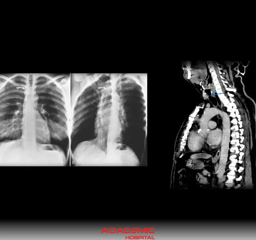
What Is Tracheal Radiology? Tracheal CT and Imaging Methods
Contents
- What Is the Trachea?
- What Is Tracheal Radiology?
- Why Is Tracheal Imaging Performed?
- What Are the Symptoms of Tracheal Diseases?
- Which Methods Are Used in Tracheal Radiology?
- What Is Tracheal CT?
- How Is Tracheal Radiology Performed?
- The Importance of Early Diagnosis in Tracheal Diseases
- Frequently Asked Questions
- References
The trachea, also known as the windpipe, is an important part of the respiratory system. Diseases or narrowing in this structure, which allows us to breathe in and out, can lead to serious breathing problems. Tracheal radiology refers to radiological evaluations that allow the windpipe to be visualized.
With these imaging studies, narrowing, masses, infections or structural problems in the trachea can be examined in detail.
Get information about tracheal imaging and radiological evaluation.
Book an AppointmentWhat Is the Trachea?
The trachea is the structure that allows air to reach the lungs. It begins below the larynx and extends down to the bronchi.
Conditions that may develop in the trachea include:
- Narrowing,
- Obstruction,
- Tumors,
- Infections,
- Trauma,
and these may cause respiratory problems.
What Is Tracheal Radiology?
Tracheal radiology includes radiological examinations performed to visualize the windpipe. The purpose is to evaluate the structure of the trachea and detect possible diseases.
Through these examinations, the following can be evaluated:
- Narrowing of the windpipe,
- Masses or tumors,
- Airway obstruction,
- Congenital structural problems,
- Trauma-related damage.
Why Is Tracheal Imaging Performed?
Doctors may request tracheal imaging in the following situations:
- Shortness of breath,
- Long-lasting cough,
- Wheezing,
- A sound during breathing,
- Recurrent lung infections,
- Difficulty swallowing,
- Airway evaluation after trauma.
In some cases, a patient’s complaints may resemble other diseases, so detailed imaging may be needed.
What Are the Symptoms of Tracheal Diseases?
Symptoms that may occur in tracheal diseases include:
- Difficulty breathing,
- Wheezing,
- Chronic cough,
- Voice changes,
- A feeling of pressure in the chest,
- Shortness of breath during exercise.
In advanced cases, severe airway narrowing may develop.
Which Methods Are Used in Tracheal Radiology?
Different imaging methods may be used to evaluate the trachea.
Chest X-ray
It may be used in initial assessments. Some major airway problems may be visible on X-ray.
Computed Tomography (CT)
It provides detailed imaging of the trachea. It is frequently preferred especially for evaluating narrowing, masses or structural abnormalities.
Magnetic Resonance Imaging (MRI)
In some special cases, it may be used to evaluate soft tissues.
Fluoroscopy
It may be preferred for dynamic evaluation of the airway.
What Is Tracheal CT?
Tracheal CT is an advanced imaging method that provides detailed cross-sectional visualization of the windpipe.
It plays an important role especially in evaluating:
- Tracheal narrowing,
- Tumors,
- Compressing structures,
- Trauma,
- Infections.
In some cases, contrast material may be used for a more detailed examination.
How Is Tracheal Radiology Performed?
The process may vary depending on the imaging method to be performed.
During CT examinations, the patient lies inside the scanner and is asked to remain still for a short time. The procedure is usually painless and brief.
If contrast-enhanced imaging is planned, some preparations may be required before the procedure.
Conditions you should report before the procedure:
- Pregnancy or suspected pregnancy
- Known contrast material allergy
- Kidney disease or history of having a single kidney
- Previous surgery or trauma history
- Regularly used medications
The Importance of Early Diagnosis in Tracheal Diseases
Tracheal diseases may go unnoticed for a long time in some people. Progressive airway narrowing in particular may cause serious breathing problems.
With early diagnosis:
- Treatment planning can be done more quickly,
- Breathing problems can be brought under control,
- Possible complications can be prevented.
It is important to consult a doctor in cases of long-lasting shortness of breath or unexplained cough.
Get Information About Tracheal Imaging Procedures
You can get information from Academic Hospital specialists for shortness of breath, wheezing, chronic cough and airway evaluation.
Frequently Asked Questions
References
The general information in this article about tracheal diseases, tracheal narrowing, imaging methods and contrast material safety is supported by the following reliable health sources.
- Cleveland Clinic, “Tracheal Stenosis: Symptoms, Causes & Treatment”, clevelandclinic.org
- Cleveland Clinic, “Tracheomalacia: Symptoms, Causes & Treatment”, clevelandclinic.org
- RadiologyInfo.org, “Patient Safety: Contrast Material”, radiologyinfo.org
- RadiologyInfo.org, “Patient Safety”, radiologyinfo.org