What Is Charcot Arthropathy?
- Swelling of the foot,
- Redness on the skin,
- Increased temperature in the foot,
- When symptoms such as slight difficulty in walking appear, a specialist doctor must be consulted. If noticed at an early stage, the treatment process becomes more successful.
- Deformation in the shape of the foot (collapsed foot appearance),
- Loss of balance and difficulty walking,
- Pressure sores and easily opening chronic wounds under the foot,
- Color changes and deformities on the skin.
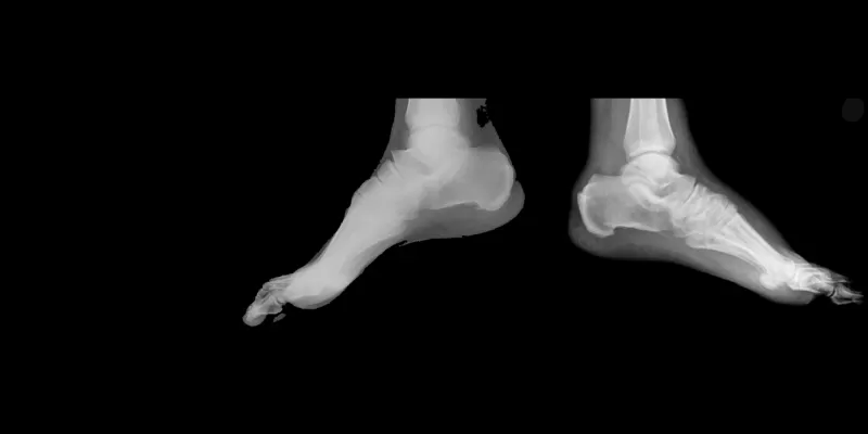
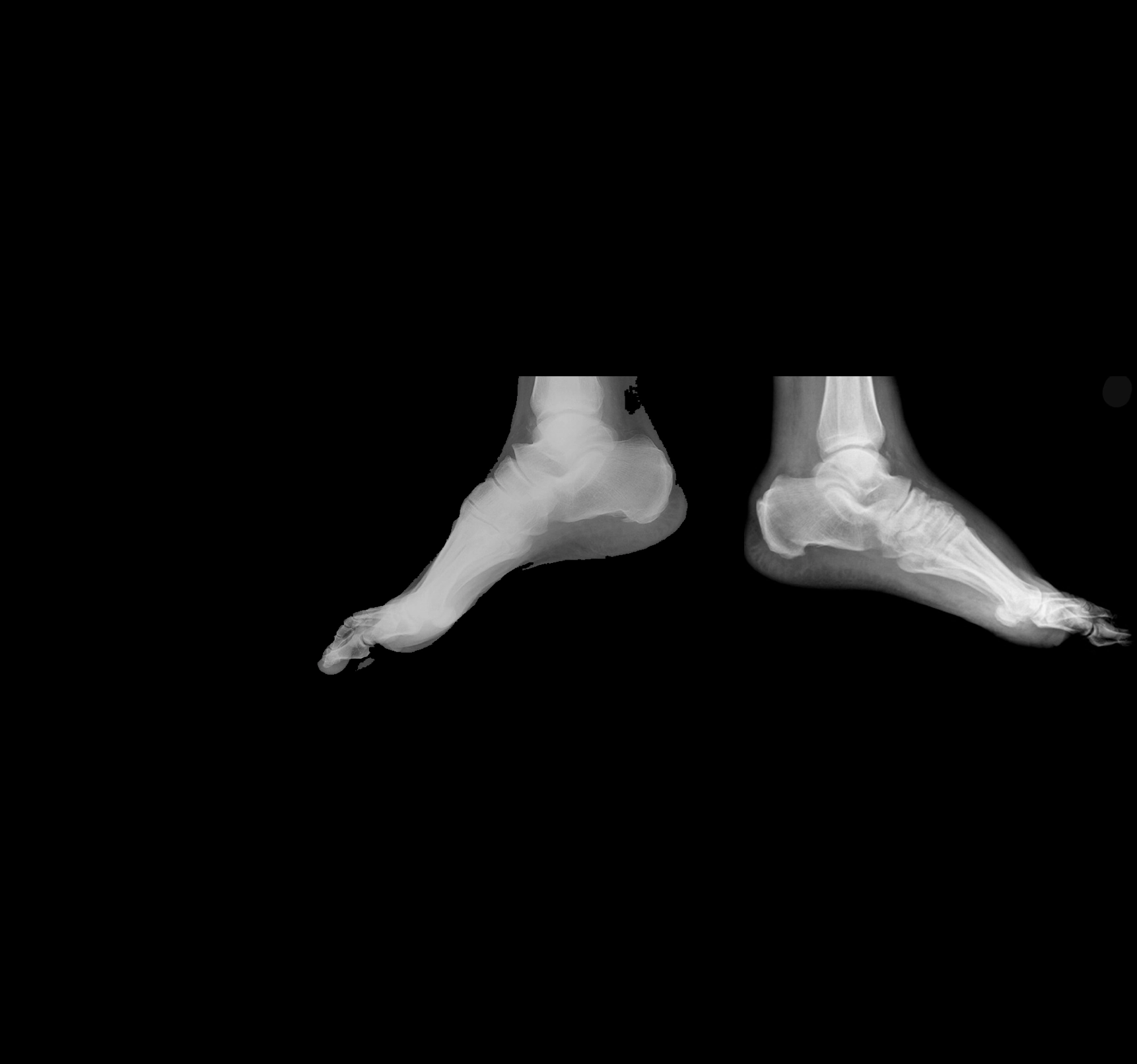
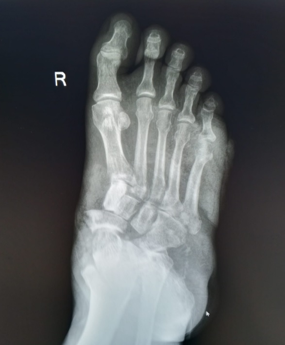
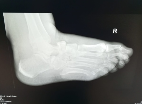
- X-ray: It can show bone fractures, joint space narrowings, and deformities in the foot and ankle.
- Magnetic Resonance (MRI): It enables the visualization of bone marrow edema and small fractures in the early period.
- Computed Tomography (CT): It reveals detailed structural breakdowns in the joints.
Also known as "Neuropathic Arthropathy", it is a serious orthopedic problem characterized by progressive bone and joint damage in the foot and ankle.
It occurs as a result of desensitization in the joints due to nerve damage and the absence of pain sensation despite loading.
What Are the Symptoms of Charcot Arthropathy?
In its initial stages, it generally presents with inflammation-like findings. Due to nerve damage, patients usually do not feel pain or feel it very mildly.
In its advanced stages, serious structural deformations occur in the joints. In this stage, symptoms become more distinct. During this period, since permanent damage may form in the foot, treatment is more difficult and sometimes surgical intervention may be required.
 WhatsApp Info Line
 How Is Charcot Arthropathy Diagnosed?
When its diagnosis is made at an early stage, it carries great importance in terms of stopping the progression of the disease and preventing serious deformities. However, since this disease is frequently confused with infections or vascular diseases, delays can be experienced in diagnosis.
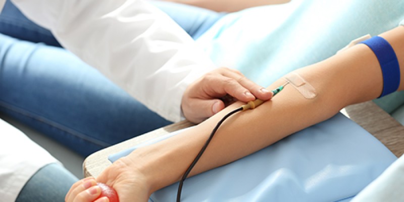
The diagnosis process usually begins with a detailed physical examination. The doctor evaluates the swelling, redness, temperature difference, and deformity in the foot. The patient's history of diabetes and neuropathy status also provide important clues in diagnosis. Since it is generally confused with infection, blood tests are also used in diagnosis.
Different imaging methods are utilized to confirm the diagnosis.
Who Gets Charcot Arthropathy? Risk Group and Risk Factors
The probability of seeing the disease is not the same in everyone; some individuals are located in the risk group and can encounter this disease at a higher rate.
Individuals who are most frequently seen among diabetes patients, especially who have developed diabetic neuropathy, meaning those who experience loss of sensation in their feet, constitute the most important risk group of this disease. Charcot joint disease occurs more frequently in people whose blood sugar control is irregular and who have been living with diabetes for a long time.
Different health problems that cause peripheral neuropathy can also increase the risk. Alcohol use, nerve injuries, and neurological diseases such as multiple sclerosis (MS) are among the important risk factors outside of diabetes. In addition, trauma, sprains, or fractures previously sustained to the foot, circulatory disorders, and smoking also facilitate the development of Charcot arthropathy.
The probability of seeing the disease is higher in individuals over 40 years old. Because over time, both nerve damage and the weakening in the joint structure become distinct. The risk is much higher especially in patients whose diabetes duration is 10 years and above, and who have a prior history of diabetic foot. For these individuals, having regular foot examinations and taking early symptoms into consideration carries critical importance to prevent the progression of the disease.
What Are the Treatment Methods for Charcot Arthropathy?
Treatment is primarily initiated by bringing diabetes under control. In this process, weight loss and diet constitute the first-step approach. In the second step, when disease findings appear, putting weight on the foot is eliminated through the use of crutches or a walker. This process must continue until the existing wounds on the foot heal, the swelling and redness resolve, and the joint becomes able to bear weight again. In patients without wounds, appropriate orthopedic shoe planning is made.
The treatment of formed open wounds, the elimination and relief of swelling in the joint are provided. Along with the progression of deformities in advanced stages, joint structures in the foot and ankle break down and the obligation to bear weight arises. In this case; starting from orthopedic surgeries aimed at fusing large joints, surgical procedures extending to partial foot or below-knee amputations are planned in cases deemed necessary. The goal is to ensure that the foot becomes a functional organ capable of bearing the body again.
Treatment initiated at an early stage plays a critical role in preventing serious complications that may form in advanced stages.

Op. Dr. Sinan Kılıç
Orthopedics and Traumatology
You can click here for appointments and detailed information.