

 WhatsApp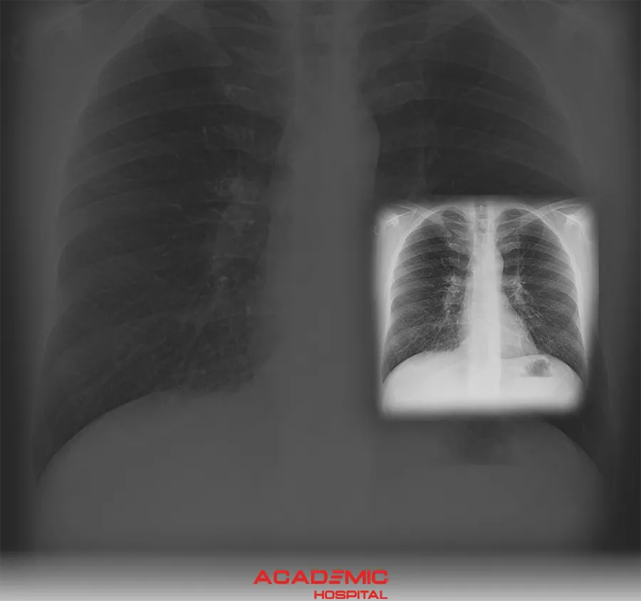
What Is a Chest X-ray? When Is It Performed and Is It Harmful?
Contents
What Is a Chest X-ray?
A chest X-ray is a fast and commonly used radiographic imaging method that helps evaluate the lungs, heart, airways, chest wall, diaphragm and spine bones. In medical terminology, it is also called chest radiography.
What Is a Chest X-ray Used For?
A chest X-ray may be used to evaluate many conditions such as lung infections, suspected mass or nodule, fluid accumulation, rib fractures and heart enlargement.
With images obtained in a short time, physicians can gather important information about the respiratory system and chest area. In some cases, a chest X-ray alone may not be sufficient; if needed, the physician may request computed tomography (CT), ultrasound, laboratory tests or further evaluation.
You can contact us for evaluation if you have persistent cough, shortness of breath, chest pain or respiratory infection symptoms.
Book an AppointmentWhen Is a Chest X-ray Performed?
A chest X-ray may be one of the first diagnostic tools used in complaints such as persistent cough or sputum, shortness of breath and chest pain.
- Febrile respiratory infections such as pneumonia or bronchitis
- Tuberculosis screening or suspicion
- Suspicion of a lung mass, nodule or tumor
- Suspicion of rib fracture, pneumothorax or internal bleeding after chest trauma
- Preoperative assessment when considered necessary by the physician
- Physician-recommended assessment in individuals who smoke, based on symptoms or risk factors
- Evaluation of findings such as heart enlargement or pleural fluid
When considered necessary by the physician, chest X-ray plays an important role in early diagnosis and treatment planning.
How Is a Chest X-ray Performed?
A chest X-ray usually takes a few minutes. The patient stands in front of a special X-ray device, is positioned by the technologist and holds their breath briefly. No pain or discomfort is usually felt during the procedure.
Before the exam, metal jewelry, necklaces or clothing accessories that may affect the chest area may need to be removed. Following the technologist’s positioning and breath-holding instructions is important for image clarity.
Is a Chest X-ray Harmful?
Because a chest X-ray uses X-rays, it involves a low dose of radiation. When it is not repeated unnecessarily and is considered medically necessary by a physician, its diagnostic benefit is generally greater than the low radiation risk. Pregnancy or suspected pregnancy should be reported to the doctor and radiology team.
A chest X-ray is an important imaging method in the evaluation of lung and chest diseases. Having it performed when recommended by a physician is a simple but important diagnostic step for your health.
The Need for a Chest X-ray Should Be Evaluated by a Physician
In cough, shortness of breath, chest pain, trauma or preoperative assessment, the appropriate imaging method is planned individually.
Frequently Asked Questions
References
- RadiologyInfo. Chest X-ray (Radiography)
- RadiologyInfo. Radiation Dose from X-Ray and CT Exams
- FDA. Medical X-ray Imaging
- FDA. Radiography