

 WhatsApp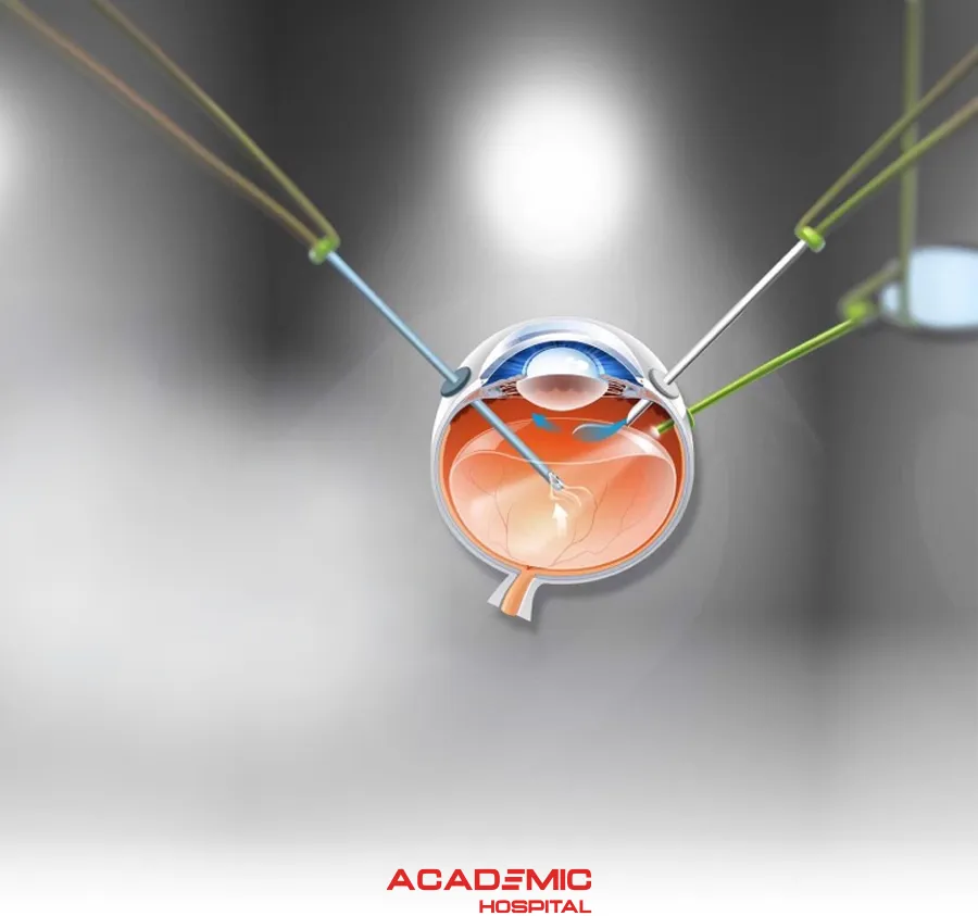
What Is Vitreoretinal Surgery? Procedures, Indications and Recovery
Contents
- What Is Vitreoretinal Surgery?
- Which Techniques Are Used?
- What Is Vitrectomy?
- Which Conditions May Require Surgery?
- Retinal Detachment Surgery
- Macular Hole and Epiretinal Membrane
- Surgery for Diabetic Retinopathy
- Is Surgery Used for Retinal Vein Occlusion?
- Why Are Gas or Silicone Oil Used?
- What Are the Risks?
- What Happens Before Surgery?
- What Happens After Surgery?
- Can You Fly With a Gas Bubble in the Eye?
- Which Symptoms Require Urgent Assessment?
- Frequently Asked Questions
What Is Vitreoretinal Surgery?
Vitreoretinal surgery refers to microsurgical procedures used to treat diseases affecting the retina and vitreous in the posterior part of the eye.
The retina is the light-sensitive tissue lining the back of the eye. The vitreous is the clear gel that fills much of the inside of the eye.
Vitreoretinal surgery may be required for conditions such as retinal detachment, macular hole, epiretinal membrane, vitreous haemorrhage and selected complications of advanced diabetic eye disease.
Which Techniques Are Used in Vitreoretinal Surgery?
The procedure is selected according to the type, location and severity of the retinal condition.
Techniques can include:
- Pars plana vitrectomy
- Endolaser treatment
- Membrane peeling
- Scleral buckling
- Cryotherapy
- Gas or silicone-oil tamponade
What Is Vitrectomy?
Pars plana vitrectomy is a microsurgical procedure in which the vitreous gel is removed to provide access to the retina and allow other retinal procedures to be performed.
Depending on the condition, surgery can involve clearing vitreous haemorrhage, removing scar tissue or membranes, draining subretinal fluid, applying laser to retinal tears or placing a gas bubble or silicone oil inside the eye.
What Is a Scleral Buckle?
During scleral buckling, a silicone band or supporting element is placed around or on the outside of the eye.
The purpose is not simply to “strengthen the sclera”. The buckle brings the eye wall closer to the retinal break and helps reduce vitreoretinal traction so that the break can seal.
Which Conditions May Require Vitreoretinal Surgery?
- Rhegmatogenous retinal detachment
- Tractional or complex retinal detachment
- Macular hole
- Epiretinal membrane
- Vitreomacular traction
- Non-clearing vitreous haemorrhage
- Selected complications of proliferative diabetic retinopathy
- Dislocated lens material or selected intraocular-lens complications
- Certain intraocular foreign bodies
- Selected severe intraocular infections
Not every retinal disease requires surgery. Observation, laser treatment or intravitreal injections may be appropriate for many conditions.
How Is Retinal Detachment Treated Surgically?
Retinal detachment occurs when the retina separates from the wall of the eye.
The objectives of treatment are to return the retina to its anatomical position, seal retinal tears and prevent additional fluid from passing underneath the retina.
Depending on the type of detachment, treatment can include vitrectomy, scleral buckling, pneumatic retinopexy or a combination of techniques.
Laser or cryotherapy may be applied around retinal tears to create a permanent seal.
Surgery for Macular Hole and Epiretinal Membrane
Macular Hole
A full-thickness macular hole can cause blurred or distorted central vision.
When surgery is required, vitrectomy is commonly performed to relieve traction on the macula, and a temporary gas bubble is often placed inside the eye.
A specific postoperative head position may be recommended depending on the characteristics of the hole and the surgical technique.
Epiretinal Membrane
An epiretinal membrane is a thin fibrocellular membrane that can develop on the surface of the retina, particularly over the macula.
Not every epiretinal membrane requires surgery. Vitrectomy and membrane peeling may be considered when there is meaningful loss of visual acuity, visual distortion or impairment of daily visual function.
When Is Surgery Used for Diabetic Retinopathy?
Surgery is not required at every stage of diabetic retinopathy.
In advanced proliferative diabetic retinopathy, abnormal blood vessels can bleed and fibrous tissue can pull on the retina.
Vitrectomy may be required for situations such as:
- Dense vitreous haemorrhage that does not clear
- Tractional retinal detachment threatening or involving the macula
- Significant fibrovascular traction on the retina
Is Vitreoretinal Surgery Used for Retinal Vein Occlusion?
Standard treatment for retinal vein occlusion does not involve surgically opening the blocked vein to restore normal blood flow.
Macular oedema associated with retinal vein occlusion is generally treated with intravitreal anti-VEGF therapy. Steroid treatment or laser may be considered in selected cases.
Vitrectomy is more likely to be used for complications such as non-clearing vitreous haemorrhage or other associated vitreoretinal pathology rather than to reverse the vascular occlusion itself.
Why Are Gas or Silicone Oil Used?
Gas or silicone oil can be placed inside the eye to support the retina internally while healing occurs.
A gas bubble gradually becomes smaller and is naturally replaced by the eye's own fluid.
Silicone oil may be used for longer-term tamponade in selected complex retinal detachments and may later require a separate procedure for removal.
What Are the Risks of Vitreoretinal Surgery?
Possible complications include:
- Intraocular infection
- Bleeding
- New retinal tear or retinal detachment
- Raised or reduced intraocular pressure
- Progression of cataract
- Recurrent membrane formation
- Persistence or reopening of a macular hole
- Failure to achieve the expected improvement in vision
- Rarely, permanent loss of vision
The individual risk depends on the operation performed and the condition of the eye before surgery.
What Happens Before Vitreoretinal Surgery?
A detailed retinal examination is performed before a decision for surgery is made.
Investigations can include dilated retinal examination, optical coherence tomography (OCT), retinal photography and B-scan ultrasonography when the retina cannot be visualised directly.
OCT is particularly important in the assessment and surgical planning of macular hole, epiretinal membrane and vitreomacular traction.
What Happens After Vitreoretinal Surgery?
Recovery depends on the condition and procedure performed.
Temporary blurred vision, mild irritation and redness may occur during the early postoperative period.
Prescribed eye drops should be used as directed and postoperative retinal examinations should not be missed.
If a gas bubble has been placed, vision may remain significantly blurred until the bubble becomes smaller.
A specific head position may be required after certain operations. The required position and its duration vary between patients.
Can You Fly With a Gas Bubble in the Eye?
No. Air travel and significant increases in altitude must be avoided while an intraocular gas bubble remains in the eye. Lower atmospheric pressure can cause the gas to expand and dangerously increase intraocular pressure.
Patients with an intraocular gas bubble must also inform any anaesthesia team before another procedure. Nitrous oxide anaesthesia can expand intraocular gas and cause serious complications.
The retinal surgeon should confirm when the gas has completely resolved and when air travel is safe again.
Which Symptoms Require Urgent Assessment After Surgery?
- Sudden or marked loss of vision
- Severe or increasing eye pain
- Markedly increasing redness or swelling
- Purulent discharge
- New flashes or a sudden increase in floaters
- A curtain or shadow in the visual field
- Eye pain accompanied by severe nausea or vomiting
Retinal Treatment Is Individualised
Whether vitrectomy, scleral buckling, laser, intravitreal treatment or observation is appropriate depends on the retinal condition and the individual eye.
Frequently Asked Questions
References
- Academic Hospital. Vitreoretinal Surgery
- American Society of Retina Specialists. Vitrectomy
- American Society of Retina Specialists. Retinal Detachment
- American Society of Retina Specialists. Macular Hole
- American Society of Retina Specialists. Epiretinal Membranes
- American Society of Retina Specialists. Branch Retinal Vein Occlusion