

 WhatsApp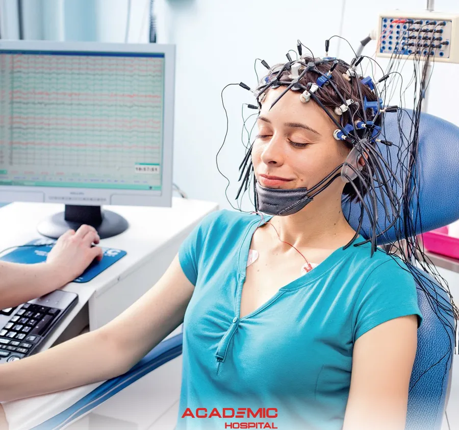
What Is an EEG (Electroencephalography)? How Is It Performed and When Is It Used?
Contents
- What Is an EEG?
- What Does an EEG Show?
- How Is an EEG Performed?
- How Long Does an EEG Take?
- When Is an EEG Used?
- What Is the Role of EEG in Epilepsy?
- Does a Normal EEG Exclude Epilepsy?
- What Are the Different Types of EEG?
- What Is a Sleep or Sleep-Deprived EEG?
- What Is Video EEG?
- How Should You Prepare?
- What Activation Procedures May Be Used?
- Is an EEG Painful or Harmful?
- EEG at Academic Hospital
- Frequently Asked Questions
What Is an EEG?
Electroencephalography (EEG) is a non-invasive neurophysiological test that records electrical activity generated by the brain using electrodes placed on the scalp.
The electrical activity generated by brain cells is detected at the scalp and displayed as waveforms on a computer. The resulting recording is called an electroencephalogram.
EEG is particularly important in the assessment of suspected epileptic seizures, classification of seizure types or epilepsy syndromes and selected disorders of brain function.
What Does an EEG Show?
An EEG allows specialists to evaluate the rhythm, frequency, distribution and characteristic patterns of electrical brain activity.
The recording may demonstrate:
- Normal activity appropriate for age and state of alertness
- Epileptiform discharges
- Focal or generalised slowing
- Changes associated with sleep
- Electrical changes occurring during a clinical event
EEG findings should be interpreted together with the patient's symptoms, neurological examination and other investigations where appropriate.
How Is an EEG Performed?
Small electrodes are positioned at defined locations on the scalp. They are usually attached using electrode paste, gel or an electrode cap.
The electrodes do not send electricity into the brain. They only detect electrical activity that can be measured at the scalp.
The patient is asked to remain as relaxed and still as possible. Depending on the purpose of the test, periods with the eyes open and closed, wakefulness and sleep may be recorded.
The EEG is recorded by appropriately trained neurophysiology staff and interpreted by a specialist in conjunction with the relevant clinical information.
How Long Does an EEG Take?
The duration depends on the type and clinical purpose of the EEG.
Current IFCN and ILAE minimum recording standards suggest, excluding preparation time:
- At least 20 minutes of technically satisfactory recording for a routine EEG
- At least 30 minutes for a sleep EEG
The overall appointment is longer because electrode preparation, application and removal also take time. NHS patient guidance notes that a standard EEG appointment commonly takes around one hour in total.
Ambulatory and prolonged video EEG recordings may continue for many hours or several days depending on clinical need.
When Is an EEG Used?
EEG is most commonly used in the assessment of seizures and suspected epilepsy.
It may be requested to:
- Assess events suspected to be epileptic seizures
- Help classify seizure type or epilepsy syndrome
- Investigate recurrent seizure-like events
- Assess selected unexplained episodes of altered awareness or consciousness
- Evaluate certain encephalopathies and disorders of brain function
- Assess selected sleep-related events that may resemble seizures
- Correlate clinical events with electrical activity during prolonged video EEG monitoring
What Is the Role of EEG in Epilepsy?
Epilepsy is fundamentally a clinical diagnosis. The description of the event, witness information and neurological assessment are central to diagnosis.
When an epileptic seizure is suspected, EEG can support the diagnosis and provide information about seizure type or epilepsy syndrome.
NICE recommends considering a routine awake EEG when the clinical history and examination suggest an epileptic seizure.
If EEG is requested after a first suspected seizure, performing it promptly may improve diagnostic yield. NICE recommends that it is ideally performed within 72 hours of the seizure.
Does a Normal EEG Exclude Epilepsy?
No.
A person with epilepsy may have a completely normal routine EEG because the test records electrical activity only during the recording period.
A normal EEG therefore must not be used by itself to exclude a diagnosis of epilepsy.
Similarly, not every EEG abnormality means that a person has epilepsy. Findings need to be interpreted in the context of the clinical history.
What Are the Different Types of EEG?
- Routine EEG: A standard-duration EEG recording.
- Sleep EEG: Includes recording during sleep.
- Sleep-deprived EEG: Performed after a prescribed reduction in sleep before the examination.
- Ambulatory EEG: Uses a portable recorder to monitor electrical activity over a longer period during usual daily activities.
- Video EEG: Records video and EEG simultaneously.
What Is a Sleep or Sleep-Deprived EEG?
Some epileptiform abnormalities are more likely to appear during sleep. A sleep EEG may therefore be considered when routine EEG has not provided sufficient information or when the suspected events are related to sleep.
For a sleep-deprived EEG, the patient is instructed to reduce sleep before the examination. This can make sleep during the recording more likely and can increase the likelihood of detecting certain epileptiform abnormalities.
Because sleep deprivation can affect seizure susceptibility, patients should follow the specific instructions provided by the EEG service rather than deciding independently how much sleep to miss.
What Is Video EEG?
Video EEG records the patient's behaviour and movements at the same time as the EEG.
This allows clinicians to compare what happens clinically during an event with any simultaneous electrical changes in the brain.
Prolonged video EEG monitoring can be useful in distinguishing epileptic seizures from non-epileptic events and for detailed assessment of selected people with epilepsy.
How Should You Prepare for an EEG?
Instructions vary according to the type of EEG.
For a routine EEG, patients are generally advised to:
- Wash their hair and keep it clean
- Avoid hair gel, oil, mousse, spray and similar products
- Eat normally and avoid skipping meals unless specifically instructed otherwise
- Tell the clinical team about all medicines being taken
- Not stop regular medication unless specifically instructed by a clinician
- Follow the EEG service's sleep instructions when a sleep-deprived EEG is planned
What Activation Procedures May Be Used During EEG?
Activation procedures may be used to assess electrical brain activity under different conditions.
These can include:
- Opening and closing the eyes
- Hyperventilation: breathing more deeply and rapidly for a defined period
- Intermittent photic stimulation: exposure to flashing lights at different frequencies
Not every procedure is appropriate for every patient. The potential benefits and risks are considered according to the clinical situation.
Hyperventilation or flashing lights can rarely provoke a seizure in susceptible individuals, which is why these procedures are carried out in a controlled clinical setting.
Is an EEG Painful or Harmful?
A routine EEG is non-invasive and generally painless.
The electrodes only record electrical activity and do not deliver electricity into the brain or body.
Occasionally, mild temporary redness or skin irritation may occur at an electrode site.
Sleep deprivation, hyperventilation and photic stimulation carry a small risk of provoking a seizure in susceptible individuals. Whether these methods are used is determined according to the clinical situation.
EEG at Academic Hospital
According to the Academic Hospital Neurology service, routine EEG and sleep/wake EEG recordings are available according to clinical need.
The appropriate type of EEG is selected according to the patient's symptoms, event history and neurological assessment.
EEG Results Must Be Interpreted in Clinical Context
EEG is an important investigation in the assessment of seizures and epilepsy, but a normal EEG does not exclude epilepsy and an EEG result alone is not sufficient to establish a diagnosis.
Frequently Asked Questions
References
- Academic Hospital. What Is EEG (Electroencephalography)? Which Patient Groups Are EEGs Taken?
- Peltola ME, et al. Routine and Sleep EEG: Minimum Recording Standards of the IFCN and ILAE
- NICE. Epilepsies in Children, Young People and Adults: Diagnosis and Assessment
- NHS. EEG (Electroencephalogram)
- Cambridge University Hospitals. Routine EEG