
 WhatsApp
What Is Spina Bifida? Types, Symptoms, Diagnosis and Treatment
Contents
- What Is Spina Bifida?
- What Are the Types?
- What Is Open Spina Bifida?
- What Is Closed Spinal Dysraphism?
- Is Every Sacral Dimple Spina Bifida?
- What Are the Symptoms?
- How Is It Diagnosed?
- Can It Be Diagnosed Before Birth?
- How Is Open Spina Bifida Treated?
- Can It Be Treated Before Birth?
- What Is Tethered Cord?
- Does Closed Dysraphism Always Require Surgery?
- Long-Term Follow-Up
- Can the Risk Be Reduced?
- Frequently Asked Questions
What Is Spina Bifida?
Spina bifida is a congenital neural tube defect that develops when the neural tube, which forms the early brain and spinal cord, does not close completely during early pregnancy.
The condition can affect the vertebrae, the membranes surrounding the spinal cord, neural tissue or a combination of these structures.
Its effects vary considerably. Some people have only a small defect in the vertebral arches, while severe forms can involve exposed spinal cord and nerve tissue at birth.
What Are the Types of Spina Bifida?
- Spina bifida occulta: A small defect in the vertebral arches, usually without involvement of the spinal nerves. Many people have no symptoms.
- Meningocele: The membranes around the spinal cord protrude through an opening in the spine, generally without spinal cord tissue within the sac.
- Myelomeningocele: Both the membranes and neural tissue protrude through the spinal defect. It is one of the most severe forms.
- Myeloschisis: A severe open neural tube defect in which neural tissue remains exposed.
- Closed spinal dysraphism: A broader group including tethered cord, spinal lipomas, dermal sinus tracts and split cord malformations.
What Is Open Spina Bifida?
In open spina bifida, the spinal cord or neural tissue is not fully covered by normal skin. Myelomeningocele and myeloschisis are important examples.
Depending on the level and severity of the lesion, associated problems can include leg weakness or paralysis, sensory loss, bladder and bowel dysfunction, orthopaedic deformities, hydrocephalus and Chiari II malformation.
Neurological function varies substantially between children and depends partly on the level and extent of neural involvement.
What Is Closed Spinal Dysraphism?
Closed spinal dysraphism refers to congenital abnormalities of the spine or spinal cord in which the abnormality is covered by skin.
Examples include tethered cord, a thickened or fatty filum terminale, spinal lipoma, lipomyelomeningocele, dermal sinus tract and split cord malformation.
Some children have symptoms from birth, while others can remain asymptomatic for many years.
Is Every Sacral Dimple a Sign of Spina Bifida?
No. Small, simple sacral dimples are common in newborns and most are harmless.
Further assessment may be appropriate when a dimple is associated with features such as:
- A prominent tuft of hair
- A skin tag or tail-like structure
- A lump or lipoma
- A vascular or pigmented skin lesion
- A deep or draining opening
- An abnormal gluteal cleft
- Neurological or orthopaedic abnormalities of the legs or feet
What Are the Symptoms?
Symptoms vary according to the type and level of spinal involvement.
- Leg weakness or loss of movement
- Sensory changes in the legs
- Foot deformities
- Changes in walking
- Spinal deformity such as scoliosis
- Urinary incontinence or incomplete bladder emptying
- Bowel dysfunction
- Back or leg pain
- Progressive neurological changes during growth
How Is Spina Bifida Diagnosed?
Spina bifida can be diagnosed during pregnancy or after birth.
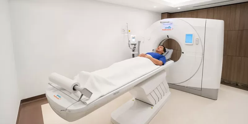
Postnatal assessment begins with physical and neurological examination. When closed spinal dysraphism is suspected, spinal ultrasound in young infants or MRI can be used depending on age and clinical findings.
MRI can provide detailed information about tethering, spinal lipomas, dermal sinus tracts, split cord malformations and other abnormalities.
Imaging findings should be interpreted alongside neurological and, when appropriate, urological findings rather than used in isolation.
Can Spina Bifida Be Diagnosed Before Birth?
Yes. Many cases of open spina bifida can be identified on prenatal ultrasound.
Evaluation can include detailed fetal ultrasound, maternal serum alpha-fetoprotein screening, fetal MRI in selected situations and additional genetic assessment when clinically indicated.
Prenatal diagnosis also allows delivery to be planned in a centre with the appropriate neonatal and neurosurgical teams.
How Is Open Spina Bifida Treated?
A newborn with an open myelomeningocele requires early assessment by neonatal and paediatric neurosurgical teams.
The exposed neural tissue should be protected from trauma and contamination while the baby's overall condition is assessed and surgical closure is planned.
The aims of closure include protecting exposed neural tissue, closing the spinal coverings appropriately, reducing infectious complications and preserving existing neurological function where possible.
Closure is generally planned during the first days of life. Current evidence does not conclusively prove that closure within 48 hours specifically lowers wound infection risk, although early surgical management remains standard clinical practice.
Can Myelomeningocele Be Treated Before Birth?
Prenatal fetal repair can be considered in selected pregnancies affected by myelomeningocele.
Evidence suggests that prenatal repair can reduce the need for cerebrospinal-fluid shunting for hydrocephalus and improve some motor outcomes in appropriately selected children.
However, fetal surgery also carries important maternal and obstetric risks, including preterm delivery, premature rupture of membranes and complications involving the uterine scar.
It is therefore considered only after detailed multidisciplinary assessment in experienced fetal-surgery centres.
What Is Tethered Cord?
Tethered cord occurs when the spinal cord is abnormally attached within the spinal canal and cannot move freely.
As a child grows, increased tension on the cord can in some patients lead to new or progressive leg weakness, sensory change, pain, changes in walking, worsening foot deformity or scoliosis, and deterioration in bladder or bowel function.
Assessment can involve neurological examination, MRI and, when appropriate, urodynamic evaluation.
Does Closed Spinal Dysraphism Always Require Surgery?
No. Not every closed spinal dysraphism automatically requires surgery.
The decision depends on the type of abnormality, neurological findings, bladder and bowel function, imaging findings, age and whether symptoms are progressing.
In symptomatic tethered cord syndrome, surgical untethering can be considered to preserve neurological function. In completely asymptomatic patients, the balance between prophylactic surgery and surveillance is more complex and remains an area of clinical debate.
Other abnormalities, such as a dermal sinus tract with potential infection risk, may require a different management strategy.
How Is Spina Bifida Followed Long Term?
Management of myelomeningocele extends far beyond surgical closure of the back.
Long-term care can involve paediatric neurosurgery, paediatric urology, orthopaedics, rehabilitation medicine, nephrology, paediatrics and physiotherapy.
Hydrocephalus, Chiari II malformation, tethered cord, bladder and kidney function, musculoskeletal development and mobility should be monitored throughout childhood and, when appropriate, adulthood.
Can the Risk of Spina Bifida Be Reduced?
Not all cases of spina bifida are preventable. However, adequate folic acid before conception and during early pregnancy reduces the risk of neural tube defects.
The CDC recommends that all women who can become pregnant consume 400 micrograms of folic acid every day.
People who have previously had a pregnancy affected by a neural tube defect may require a different dose and should receive individual medical advice before planning another pregnancy.
Treatment Depends on the Type of Spina Bifida
Open myelomeningocele requires prompt neonatal neurosurgical assessment, while closed spinal dysraphism should be managed according to anatomy, neurological findings and bladder and bowel function.
Frequently Asked Questions
References
- Academic Hospital. Spina Bifida
- Centers for Disease Control and Prevention. About Spina Bifida
- Centers for Disease Control and Prevention. Neural Tube Defects
- Spina Bifida Association. Neurosurgery Guideline
- Congress of Neurological Surgeons. Closure of Myelomeningocele Within 48 Hours to Decrease Infection Risk
- Agency for Healthcare Research and Quality / NCBI. Diagnosis and Treatment of Tethered Spinal Cord
- Centers for Disease Control and Prevention. Folic Acid: Sources and Recommended Intake